Header Image - Gluten Light

Tag Archives

2 Articles

A recent and in-depth research regarding the influence of gut microbiota, diet and exercise on intestinal permeability II part

by luciano

Link I part A recent and in-depth research regarding the influence of gut microbiota, diet and exercise on intestinal permeability. I Part

6 Exercise as a regulator of intestinal barrier integrity

Regular moderate physical exercises are one of the most common recommendations for the prevention of various pathologies, including disruption of the integrity of the intestinal barrier. This may be due to the influence of the gut microbiota. In particular, exercises have been found to increase gut bacterial diversity (Hintikka et al., 2023). However, effects of physical exercises depend on their intensity. For example, endurance athletes have a high incidence of gastrointestinal disorders and the “leaky” gut is one of the most common disorders (Ribeiro et al., 2021). It is characterized by dysfunction of the intestinal epithelial barrier and its excessive permeability. This results in penetration of harmful microorganisms, toxins or undigested food particles into the bloodstream and has a negative effect on health of the whole organism (Aleman et al., 2023).
The effect of exercise on intestinal permeability depends on its duration and intensity. For example, people who exercise frequently and intensely have the same mortality rates as people who lead a sedentary lifestyle (Van Houten et al., 2015). A 60 min bout of intensive treadmill running increased the permeability of the small intestine in runners, whereas low-intensity running had no such effect (Pals et al., 1997). Using the overtraining model with male C57BL/6 mice, it was established that exhaustive exercise exacerbated intestinal inflammation, disrupted integrity and enhanced intestine wall permeability (Hou et al., 2020). Sustained strenuous exercise in racing sled dogs increased the intestinal permeability and the frequency of gastric erosions or ulcerations (Davis et al., 2005). High-intensity interval running increased intestine wall permeability and intestinal-fatty acid binding protein (I-FABP) release in male runners (Pugh et al., 2017). I-FABP is a cytoplasmic protein expressed exclusively in the enterocytes of the small intestine and its increased concentration in the blood is used as a marker of damage to intestinal epithelial cells (Sikora et al., 2019).
Physical exercise of low/moderate intensity can often have positive effects and can be considered as a method of non-pharmacological intervention in inflammatory bowel disease (Ordille and Phadtare, 2023). For example, mice that swam for 30 min before inducing intestinal barrier dysfunction had less intestinal dysfunction compared to mice that had not swum before. This might happen due to a strengthening of antimicrobial function of the intestine as a result of the increase in expression of antimicrobial peptides (Luo et al., 2014). Obese mice that were trained on a motorized treadmill for 45 min per day 5 days a week for 12 weeks had higher expression levels of colonic ZO-1 and occludin. Moderate exercise effectively prevented the development of dysbacteriosis caused by the HFD, as well as intestinal pathology (Wang et al., 2022). Dysbacteriosis and impaired intestinal barrier integrity induced by HFD in wild type mice was prevented by exercise. Exercise on a motor-driven rodent treadmill for 5 days a week for a total of 15 weeks significantly reversed the pathological changes. Ablation of Sestrin 2 protein attenuated the protective effects of exercise, suggesting its involvement in regulation of intestinal permeability (Yu et al., 2022). Thus, it can be concluded that high-intensity exercises often have a negative effect on the integrity of the intestine, whereas low- and moderate-intensity regular exercise can have a positive effects. It may be speculated that moderate damage to the intestinal wall is a hormetic factor that may be used to train organisms to cope with severe damaging challenges. This may be used to increase the adaptive potential of organisms to prevent damaging effects of any stresses of physical and chemical nature on the integrity of the intestinal wall.

6.1 Exercise-induced heat stress

It is known that physical exertion causes heat stress and associated dysfunction of gut integrity. A systematic review examining the relationship between an exercise-induced increase in core body temperature and intestinal permeability demonstrated that the magnitude of exercise-induced hyperthermia correlated with increased intestinal permeability (Pires et al., 2017). An increase in body temperature is a signal to activate the expression of heat shock proteins (HSP) which constitutively function as molecular chaperones maintaining the native structure of the proteins. Their expression is mainly triggered by heat shock signals. During exercise, the level of HSP70 and HSP90 increase (Krüger et al., 2019). Expression of HSP is regulated at the level of heat shock factors (HSF) such as HSF1 that is expressed in all mammalian tissues. Normally it resides in the cytoplasm as a monomer. In response to stressful conditions, it trimerizes, translocates into the nucleus, binds to the heat shock element of target genes and activates the transcription of HSPs, including HSP70/90 (Noble and Shen, 2012).
In this way, exercises cause a homeostatic imbalance, while regular training is adaptive and decreases the degree of this imbalance. Potentially, a higher adaptive steady-state level of HSPs due to regular training could explain their positive effect on gut integrity. At that time, during acute physical exertion, HSPs probably cannot cope with that level of homeostatic imbalance caused exercise-induced heat stress.

6.2 Exercise-induced hypoxia
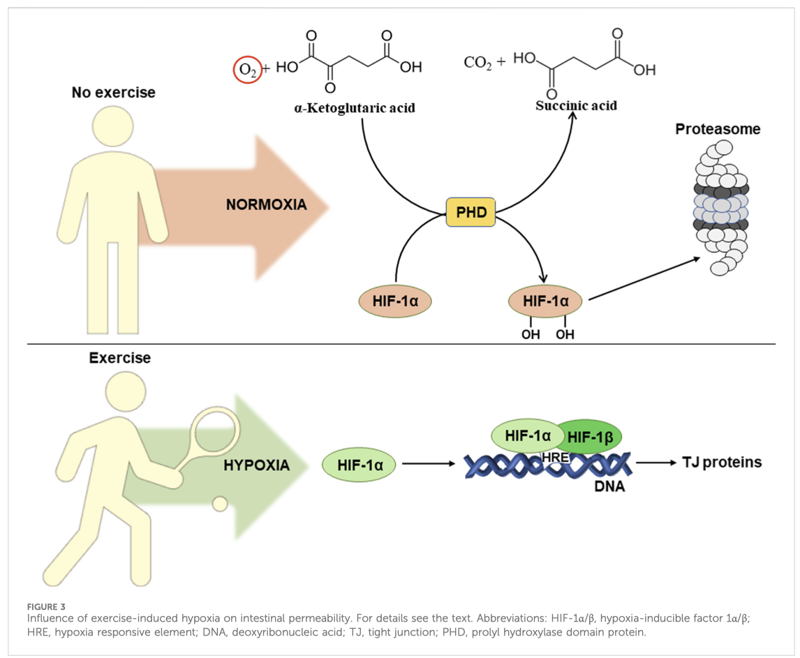
It is well known, that exercise causes a redistribution of blood flow between tissues. This leads to the development of hypoxia (decreased oxygen levels) in intestinal epithelial cells and activation of hypoxia-inducible factor alpha (HIF-1α) (Wu et al., 2020). Figure 3 schematically shows the influence of exercise-induced hypoxia on intestinal permeability. In normoxia (normal oxygen levels), prolyl hydroxylase hydroxylates HIF-1α at two proline residues (Pro 402 and Pro 564). This results in ubiquitination followed by subsequent proteasomal degradation of HIF-1α (Lee et al., 2004).

intestinal barrier

…..omissis

7 Conclusion and perspectives
The intestinal wall is a kind of checkpoint between the external and internal environments of organisms. The wall consists of three layers: mucous, epithelial, and lamina propria. The mucous layer is inhabited by microorganisms, many of which mutually beneficially coexistence within the human body. These microorganisms modulate many if not most living processes: from the development of the immune and nervous systems at early stages of life to the induction of chronic inflammation causing neurodegeneration at aging. Despite the fact that these microorganisms have coexisted with humans for many years, under certain conditions the enteral immune system of the lamina propria can perceive them as foreign and trigger a pro- inflammatory response.
Normally, the intestinal mucosa is semipermeable. It allows selective absorption of nutrients into the bloodstream but prevents the entrance of potentially harmful microorganisms and their waste products from contact with the enteral immune system. An imbalance of the intestinal microbiota, called dysbiosis, can cause a disturbance of intestinal integrity and increase intestinal permeability. Conversely, a healthy composition of the gut microbiota can contribute to the integrity of the intestinal barrier due to increased expression and induction of the assembly of TJ proteins, activation of mucus synthesis, and antioxidant action.
Disruption of intestinal barrier function may trigger development of local and even systemic inflammation.
…..omissis
In general, a vicious cycle of intestinal barrier disruption can be traced here, as excessive intestinal wall permeability provokes the development of chronic low-grade inflammation. The latter is characterized by increased production of pro-inflammatory cytokines and enhanced ROS generation, increasing intestinal barrier dysfunction.
Nutrition looks to be the simplest non-pharmacological effector of integrity and permeability of the intestinal wall. It can have both a negative effect, such as HFD inducing metabolic endotoxemia, or a positive effect, such as a diet rich in plant polyphenols or fermented dairy products, increasing the expression of TJ proteins and promoting the development of beneficial bacteria.
Exercise also can affect gut intestinal permeability. Its effects depend on duration and intensity of exercise. Acute extensive physical exertion often increases intestinal permeability which may be related to the induction of heat stress, that organisms cannot cope with at that time due to insufficient resources. On the other hand, regular low and moderate intensity exercises, that are adaptive in nature, mostly have a positive effect on the integrity of the intestine and decrease its permeability. Potentially, this may be associated with an increase in the steady-state level of HSPs and chronic activation of HIF-1α which activates the transcription of genes responsible for strengthening the intestinal barrier function.
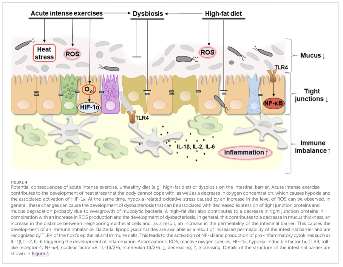
In general, it can be concluded that proper nutrition which promotes a healthy biodiversity of the gut microbiota, combined with moderate exercise, contribute to the integrity of the intestine. Disbalanced nutrition and excessive physical activity can provoke the development of dysbacteriosis and increase intestinal permeability which can potentially lead to a pro-inflammatory response. Figure 4 schematically shows potential consequences of acute intense exercises, unhealthy diet (e.g., high-fat diet), and dysbiosis on the intestinal barrier.
Taking into account all of the above, we can outline the following future prospects:
1. Development of healthy diets to support intestinal homeostasis;
2. Use of fermented dairy products as natural pre-, pro- and postbiotics to promote a healthy gut;
3. Selection of exercises to promote intestinal integrity by frequency, intensity and duration;
4. Study of the role of intestinal HIF-2α during exercise;
5. Systemic investigation of hypoxia-induced oxidative stress as a regulator of intestinal wall permeability.
Most of these perspective avenues are directed to enhance the capability of organisms to cope with disturbing factors. That increases an adaptive capability via preadaptation/hormetic mechanisms. However, some of them may be used “to patch holes” in “leaky” intestinal wall, which is characterized by increased specific permeability of the intestinal epithelium. Intestinal barrier permeability: the infuence of gut microbiota, nutrition, and exercise. Tetiana R. Dmytriv et al. DOI 10.3389/fphys.2024.1380713. PUBLISHED 08 July 2024

Note
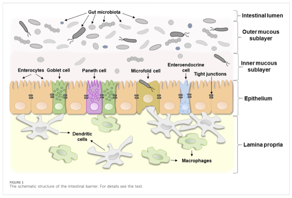
[1] The term “intestinal barrier” emphasizes the barrier function of the intestinal wall which protects organism against invading by bacteria or other microorganisms and potentially toxic components of microorganisms. In fact, it is a complex selective physical barrier that separates the internal environment of the body from the contents of the intestinal lumen (Bischoff et al., 2014). Figure 1 shows a schematic structure of the intestinal barrier. It consists of several layers: i) a mucous layer including inner and outer mucous sublayers inhabited by commensal microorganisms in a different extent, ii) a single layer of epithelial cells, and iii) the lamina propria, which consists of immune cells that instantly react to the invasion of foreign substances (Schoultz and Keita, 2020).
The first layer, the mucous layer, that consists mainly of a mesh polymer called mucin, is located on the side of the intestinal lumen. It is associated with community of commensal microorganisms, including bacteria, fungi, viruses, and parasites, that form the individual microbial community (Chelakkot et al., 2018). A change in the microbial composition that causes a sharp imbalance between beneficial and potentially pathogenic bacteria, including changes in its functional composition, metabolic activity or changes in their local distribution, is called dysbiosis or dysbacteriosis. The latter usually results from loss of beneficial bacteria, overgrowth of potentially pathogenic bacteria, or loss of overall bacterial diversity. This disrupts the homeostatic balance of the intestinal microbiota and has a negative impact on the host’s health. In particular, dysbacteriosis is implicated in a wide range of diseases (DeGruttola et al., 2016).
The second layer, the intestinal epithelium, consists of a single layer of several specialized epithelial cells, such as enterocytes, Goblet cells, Paneth cells, enteroendocrine cells, and microfold cells (Figure 1). Enterocytes form the basis of the intestinal epithelium and play a main role in the absorption of all consumed nutrients. Goblet cells constitute about 10% of specialized epithelial cells. They secrete mucus to protect the intestinal wall from digestive enzymes (Kim and Ho, 2010). Paneth cells contain secretory granules filled with antimicrobial peptides, that are secreted in low amounts constitutively and provide the antimicrobial properties of the intestinal mucosa. Under certain conditions, their secretion can increase dramatically (Yokoi et al., 2019). Enteroendocrine cells produce hormones regulating secretion of digestive enzymes and insulin, peristalsis of the intestine, satiety, and immune response (Bonis et al., 2021). Microfold cells transport bacteria and antigens from the epithelium to enteric immune cells that either activate or suppress the immune response (Jung et al., 2010). All these cell types collectively contribute significantly to gut homeostasis.
The third layer, lamina propria, is located under the epithelium and forms the enteric immune system that consists of a large number of leukocytes with macrophages and dendritic cells being the dominant cell types (Shemtov et al., 2023). Resident intestinal macrophages are located in close proximity to the gut microbiota, with which they often interact. They play a key role in immune sampling of luminal bacteria, contributing to the maintenance of intestinal homeostasis and regulated immune response.

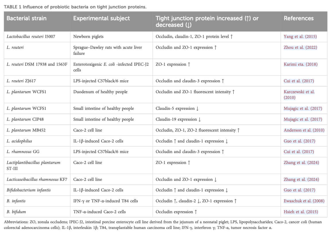
[2] TJ proteins are a complex of transmembrane and cytoplasmic proteins that form tight junctions, which seal cells together to create a selective barrier, maintain cell polarity, and regulate cell processes.

Key words
tight junction, tight junction proteins, inflammation,

A recent and in-depth research regarding the influence of gut microbiota, diet and exercise on intestinal permeability. I Part

by luciano

A recent and in-depth research regarding the influence of gut microbiota, diet and exercise on intestinal permeability.
Tetiana R. Dmytriv et al. 2024.
For the full test: Tetiana R. Dmytriv et al. DOI 10.3389/fphys.2024.1380713. PUBLISHED 08 July 2024

Highlighted
1. The intestinal wall [21consists of three layers: mucous, epithelial, and lamina propria. The mucous layer is inhabited by microorganisms, many of which mutually beneficially coexistence within the human body. These microorganisms modulate many if not most living processes: from the development of the immune and nervous systems at early stages of life to the induction of chronic inflammation causing neurodegeneration at aging. Despite the fact that these microorganisms have coexisted with humans for many years, under certain conditions the enteral immune system of the lamina propria can perceive them as foreign and trigger a pro- inflammatory response.
2. Normally, the intestinal mucosa is semipermeable. It allows selective absorption of nutrients into the bloodstream but prevents the entrance of potentially harmful microorganisms and their waste products from contact with the enteral immune system. An imbalance of the intestinal microbiota, called dysbiosis, can cause a disturbance of intestinal integrity and increase intestinal permeability.
3. Excessive intestinal wall permeability provokes the development of chronic low-grade inflammation.
4. Nutrition looks to be the simplest non-pharmacological effector of integrity and permeability of the intestinal wall. It can have both a negative effect, such as HFD inducing metabolic endotoxemia, or a positive effect, such as a diet rich in plant polyphenols or fermented dairy products, increasing the expression of TJ proteins [2] and promoting the development of beneficial bacteria.
5. Exercise also can affect gut intestinal permeability. Its effects depend on duration and intensity of exercise. Acute extensive physical exertion often increases intestinal permeability which may be related to the induction of heat stress, that organisms cannot cope with at that time due to insufficient resources. On the other hand, regular low and moderate intensity exercises, that are adaptive in nature, mostly have a positive effect on the integrity of the intestine and decrease its permeability.

“The intestinal wall is a selectively permeable barrier between the content of the intestinal lumen and the internal environment of the body. Disturbances of intestinal wall permeability can potentially lead to unwanted activation of the enteric immune system due to excessive contact with gut microbiota and its components, and the development of endotoxemia, when the level of bacterial lipopolysaccharides increases in the blood, causing chronic low-intensity inflammation. In this review, the following aspects are covered: the structure of the intestinal wall barrier; the influence of the gut microbiota on the permeability of the intestinal wall via the regulation of functioning of tight junction proteins, synthesis/degradation of mucus and antioxidant effects; the molecular mechanisms of activation of the pro-inflammatory response caused by bacterial invasion through the TLR4-induced TIRAP/MyD88 and TRAM/TRIF signaling cascades; the influence of nutrition on intestinal permeability, and the influence of exercise with an emphasis on exercise-induced heat stress and hypoxia. Overall, this review provides some insight into how to prevent excessive intestinal barrier permeability and the associated inflammatory processes involved in many if not most pathologies. Some diets and physical exercise are supposed to be non-pharmacological approaches to maintain the integrity of intestinal barrier function and provide its efficient operation. However, at an early age, the increased intestinal permeability has a hormetic effect and contributes to the development of the immune system.

Introduction
The intestinal wall is a complex system consisting of four layers: mucosa, submucosa, muscularis, and serosa. The term “intestinal barrier” emphasizes the protective component of the intestinal wall, whereas intestinal permeability is a measurable characteristic of the functional status of the intestinal barrier (Bischoff et al., 2014). The wall provides selective absorption of nutrients and other components of the intestinal lumen. At the same time, the intestinal barrier protects the body from the entrance of unwanted foreign substances, food particles, microorganisms, and their components. In normally functioning organisms, the permeability of the intestinal wall is tightly controlled but its disturbance, if not adequately fixed, can lead to many, if not most, acquired pathologies (Gieryńska et al., 2022).
The gastrointestinal tract (GIT) is inhabited by diverse microbes called gut microbiota forming very dynamic community.


Figure 1 The schematic structure of the intestinal barrier. For details see the text.

The “Old Friends Hypothesis” suggests that people coevolved with many microbes that, in addition to many physiological functions, also stimulate the development of the immune system and regulate its operation (Rook, 2023). Microbial antigens are under constant surveillance by the enteric immune system. Regulatory immune T cells are responsible for maintaining immune tolerance of homeostatic gut microbiota (Wu and Wu, 2012). However, increased intestinal permeability can promote translocation of luminal bacteria and microbial-associated molecular patterns, in particular, lipopolysaccharides (LPS) from the gut into bloodstream, triggering the development of endotoxemia and chronic low- intensity inflammation (Vanuytsel et al., 2021). Diet-induced endotoxemia is defined as metabolic endotoxemia. For example, Cani et al. (2007) established that a high-fat diet chronically increased plasma LPS concentrations two-to threefold.
Endogenous lipopolysaccharides LPS are constantly released as a result of the death of Gram-negative bacteria in the gut. At increased intestinal barrier permeability, LPS are absorbed into the portal bloodstream, from where they are transported by lipoproteins directly into the liver, forming the gut-liver axis. Further, they are metabolized by liver enzymes and excreted with bile. However, if their degradation or biliary excretion are impaired, LPS can reach the systemic circulation, where they bind to Toll-like receptor 4 (TLR4) on leukocytes, endothelial cells, and platelets, causing arterial inflammation. Ultimately, this leads to activation of blood coagulation and thrombus formation, which demonstrates that LPS-induced inflammation associated with increased intestinal wall permeability may be involved in the development of atherosclerosis and thrombotic diseases (Violi et al., 2023). In general, disruption of intestinal barrier function is involved in many GIT-related and unrelated diseases, including inflammatory bowel disease, metabolic dysfunction-associated liver disease, bile acid malabsorption, celiac disease, type I diabetes, obesity, schizophrenia, and others (Vanuytsel et al., 2021). Potentially, this could be overcome by a non-pharmacological intervention based on diet and exercises (Pražnikar et al., 2020; Ordille and Phadtare, 2023) which promote a healthy gut ecosystem and alleviate the symptoms of many pathologies.
In this review, we describe the structure of the intestinal wall and molecular mechanisms of the pro-inflammatory response caused by bacterial invasion due to the disturbance of the intestinal wall permeability, as well as influences of the gut microbiota, diet, and exercises on the permeability of the intestinal wall. Specific diets and regular low- and moderate-intensity exercises are proposed as effective non-pharmacological approaches to maintain integrity of intestinal wall and its efficient operation. However, at an early age, controlled leakage of the intestine may be necessary to trigger the development of immune system via hormetic mechanisms.

2 The structure of the intestinal barrier
…….omissis

3 Intestinal permeability
Semi-permeability or selective permeability is a crucial feature of the intestinal wall. It limits penetration of pathogens but allows the permeability of nutrients, water, and ions. Endogenous (e.g., inflammation) and exogenous (e.g., diet components, toxicants, or drugs) factors can increase intestinal permeability and cause the formation of a so-called “leaky gut.” The latter is characterized by the penetration of food antigens, commensals, or pathogenic bacteria into the blood, causing the development of inflammation (Vanuytsel et al., 2021). Some diseases can also act as a disruptor factor of the intestinal barrier. For example, several studies show that hyperglycemia, a key feature of diabetes, induces intestinal barrier dysfunction (Thaiss et al., 2018; Dubois et al., 2023). Prolonged exposure to glucose at high levels increases migration capacity of human colonic cell line Caco-2, resulting in layers appearing less organized than under physiological conditions. In particular, this is associated with decreased expression of tight junction (TJ) proteins, which contributes to the disruption of the structural network associated with them and an increase in the permeability of the intestinal barrier (Dubois et al., 2023). In turn, this contributes to the penetration of luminal bacteria, and the development of dysbacteriosis resulting in inflammation. For example, Harbison et al. (2019) showed that children with type I diabetes have gut microbiota dysbiosis associated with increased intestinal permeability. In particular, lower microbial diversity, lower numbers of anti-inflammatory bacterial species, and SCFA- producing bacteria were observed, and these changes were not explained by differences in diet. Thus, some diseases, including diabetes, can also play the role of disruptors of the intestinal barrier.
Mucus and epithelium are the most important components of the intestinal barrier that limit the development of inflammation. The mucous layer consists of two sublayers (Figure 1). The outerlayer is thick and loose. It is inhabited by a large number of commensal microorganisms that form colonies, and under healthy conditions pathogenic bacteria cannot outgrow them or penetrate further. In other words, homeostatic microorganisms efficiently compete with potentially pathogenic ones and prevent their excessive proliferation. The inner sublayer, on the contrary, is solid and contains only a few microbes (Usuda et al., 2021). The gut microbiota plays a major role in changing the composition of mucus, regulating its synthesis and degradation.
Epithelial cells are connected by TJ proteins (Lee et al., 2018) which regulate the absorption of water, ions, and dissolved substances. They include two functional categories of proteins: integral transmembrane proteins, located at the border of adjacent cell membranes, and adaptive peripheral membrane proteins that connect integral proteins with the actin cytoskeleton. The former includes occludin, claudins, junctional adhesion molecules, and tricellulin whereas the latter include zonula occludens-1 (ZO-1), ZO-2, and ZO-3 (Lee et al., 2018). The gut microbiota can influence the expression and localization of all of these TJ proteins.

3.1 Influence of the gut microbiota on tight junction proteins
TJ proteins regulate the rate of paracellular transport including the transport of consumed nutrients via the path between neighboring epithelial cells. In electron micrographs TJ proteins look like points of fusion of the membranes of neighboring cells where there is no intercellular space in these places (Gonzalez- Mariscal et al., 2003). They play the role of sensors of environmental conditions that dynamically regulate the paracellular transport of solutes (Ulluwishewa et al., 2011). Dysregulation of TJ proteins can lead to excessive permeability of the intestinal barrier.
Bacteria can change the expression and distribution of TJ proteins and thus affect intestinal permeability. For example, some pathogenic strains of Escherichia coli, including E. coli O157:H7 strain which causes bloody diarrhea, produce toxins such as Shiga toxins (STx). The latter suppress protein biosynthesis and contribute to the development of hemolytic uremic syndrome, which is a life-threatening complication. Pradhan et al. (2020) found that STx2a decreases the expression of TJ proteins such as ZO-2, occludin, and claudin-1 (Pradhan et al., 2020). However, this strain requires the presence of non-pathogenic E. coli, which enhances the expression of Stx2a. In this way, non- pathogenic E. coli decreases the expression of TJ proteins, increasing the production of the STx2a toxin by E. coli O157:H7 strain (Xiaoli et al., 2018). This indicates that, under certain conditions, even non- pathogenic microbiota can have a negative impact on intestinal wall permeability. Contrarily, the use of probiotics (living microorganisms that are beneficial to the host organism when administered in adequate amounts) may contribute to the integrity of the intestinal barrier (Ulluwishewa et al., 2011; Gou et al., 2022). In particular, Lactobacillus and Bifidobacterium species are the most commonly used probiotics. For example, Lactobacillus reuteri increases the expression of TJ proteins and thus supports the integrity of the intestinal wall (Gou et al., 2022). Oral administration of L. reuteri I5007 significantly increased the levels of claudin-1, occludin, and ZO-1 in newborn piglets. An in vitro study showed that pretreatment of intestinal porcine epithelial cell line J2 with this bacterial strain suppressed a LPS-induced decrease in TJ protein expression (Yang et al., 2015). Administration of L. plantarum into the duodenum of healthy people increased the level of ZO-1 and occludin. However, L. plantarum did not significantly affect expression of occludin in vitro human epithelial model but induced translocation of ZO-1 into the TJ region which forms a paracellular seal between epithelial cells (Karczewski et al., 2010; Caminero et al., 2023). Bifidobacterium infantis and L. acidophilus prevented dysregulation of occludin and claudin-1 levels in colon carcinoma cell line (Caco-2) stimulated by IL-1β treatment. These strains normalized their expression and contributed to the integrity of the intestinal barrier (Guo et al., 2017). For convenience, we have summarized some available information regarding the influence of different probiotic bacterial strains on TJ proteins in Table 1. In general, probiotic bacteria can both increase and decrease TJ proteins. However, in most cases, this does not cause excessive intestinal permeability, but on the contrary, normalizes it and contributes to its integrity.
Antibiotics used to treat bacterial infections may adversely affect the gut microbiota. They cause an imbalance between specific groups of bacteria and trigger the development of dysbacteriosis (Tulstrup et al., 2015). Dysbacteriosis, in turn, contributes to intestinal permeability. An increase in the population of pathogenic bacteria at dysbacteriosis which probably produce higher levels of LPS, can damage epithelial cells of the intestinal barrier and contribute to increased intestinal permeability. For example, it was shown that changes in the microbial composition correlated with an increase in intestinal permeability in alcohol- dependent subjects (Leclercq et al., 2014).
In addition, the gut microbiota is a significant source of digestive proteases used to break down host proteins for their own needs. However, excessive activity of microbial proteases can disrupt the epithelial components of the intestinal barrier due to cleavage of TJ proteins. In turn, changes in TJ proteins lead to an increase in the paracellular permeability of the epithelial barrier (Caminero et al., 2023).

3.2 The role of gut microbiota in biosynthesis and degradation of mucous layer components

omissis

3.3 Antioxidant effects of intestinal microorganisms

omissis
4 Molecular mechanisms of the activation of pro-inflammatory response caused by bacterial invasion

Dysbacteriosis of the gut microbiota can lead to disruption of intestinal barrier function and immune homeostasis. Increased intestinal permeability facilitates the translocation of microbes, their components, and microbial products into the blood stream and their recognition by the host immune cells (Longo et al., 2020). The gut microbiota is the main reservoir of pro-inflammatory endotoxins inside the body. In particular, LPS, the main component of the outer membrane of Gram-negative bacteria, can cause so-called endotoxemia. The latter develops when the level of LPS in the blood increases and this leads to the activation of a pro-inflammatory immune response triggering systemic low-grade inflammation (André et al., 2019). A diet-induced increase in LPS concentration in the blood is called metabolic endotoxemia. The level of LPS in the blood serum of mice that consumed high-fat diet (HFD) for 4 weeks is similar to its level in metabolic endotoxemia (Mohammad and Thiemermann, 2021). This clearly shows how nutrition can affect intestinal permeability and immune response.
The dynamic interaction between the gut microbiota and the intestinal immune system plays a key role in maintaining intestinal homeostasis. Host cells contain pattern recognition receptors (PRRs) which recognize bacterial pathogen-associated molecular patterns (PAMPs). The latter are highly conserved bacterial motifs, possessed in LPS, oligodeoxynucleotides, peptidoglycans, and others that can trigger host immune response (Asiamah et al., 2019).
…………omissis

4.1 Early/late activation of inflammation by TIRAP/MyD88 and TRAM/TRIF
signaling cascades

……….omissis

Some dairy products, such as kefir, have long been studied as regulators of intestinal integrity. For example, consumption of kefir for 21 days by healthy people with two washout periods in-between decreased the serum level of zonulin (Novak et al., 2020) Kefir diet also normalized zonulin level in overweight people (Pražnikar et al., 2020). Zonulin is a protein that increases the permeability of the intestinal barrier and is often involved in the development of autoimmune diseases, including type I diabetes. Zonulin causes TJ disassembly and thus violates the intestinal barrier (Fasano, 2011). Therefore, zonulin is considered a serum marker of the integrity of the intestinal wall.
Polyphenolic compounds (secondary plant metabolites with a long list of beneficial properties for humans) are other food components that improve intestinal integrity. Flavonoids are among most abundant representatives of this group. They are found mostly in fruits, vegetables, grains, tea, and wine (Kasprzak-Drozd et al., 2021). For example, the flavonoid quercetin increased intestinal integrity, as studied in Caco-2 cells (Suzuki and Hara, 2009). This effect was associated with the assembly of ZO-2, occludin and claudin-1, as well as increased expression of claudin-4 and transepithelial electrical resistance. The electrical resistance of epithelial cells is a reliable indicator of the integrity and permeability of the cell monolayer and TJ (Srinivasan et al., 2015). The consumption of quercetin in food increased the mRNA levels of occludin and ZO-1 in pigs that was accompanied by a decrease in serum endotoxin (Zou et al., 2016), a marker of metabolic endotoxemia frequently associated with increased intestinal permeability. The flavonoid kaempferol may have similar effects. In a study on Caco-2 cells during the first 6 hours after kaempferol administration, transepithelial electrical resistance increased significantly and this correlated with the assembly of occludin and claudin-3 (Suzuki et al., 2011).
A meta-analysis performed to study the effects of oral administration of phenolic compounds on the integrity of the intestinal barrier in animals confirmed their beneficial effects. In particular, the improvement of intestinal wall integrity occurs due to the three main mechanisms: i) increased expression of TJ proteins, ii) decreased levels of pro-inflammatory molecules, and iii) increased intracellular antioxidant potential (Sandoval-Ramírez et al., 2021).

………….omissis

Thus, nutrition can affect the integrity of the intestine and this is often associated with various pathological conditions. The effect mainly occurs at the level of modulation of gut microbiota composition and regulation of TJ protein operation. In this regard, healthy nutrition can be considered as a promising way to attenuate various pathologies.

A recent and in-depth research regarding the influence of gut microbiota, diet and exercise on intestinal permeability II part