Header Image - Gluten Light
Gallery

Magazine

Invecchiamento del sistema immunitario

by luciano

Panoramica delle più recenti ricerche relative all’invecchiamento del sistema immunitario e della correlazione con l’infiammazione.

In evidenza
1 – L’invecchiamento è un processo multifattoriale guidato da vari fattori intrinseci ed estrinseci, tra cui instabilità genomica, accorciamento dei telomeri (modificazioni della sequenza del DNA) [A], alterazioni epigenetiche, perdita di proteostasi, macroautofagia disabilitata, rilevamento dei nutrienti alterato [B], disfunzione mitocondriale, senescenza cellulare, esaurimento delle cellule staminali, comunicazione intercellulare alterata, infiammazione cronica e disbiosi. Questi fattori sono strettamente correlati all’invecchiamento dell’organismo e la ricerca ha dimostrato che indurli può accelerare l’invecchiamento, mentre intervenire su di essi può rallentare, arrestare o persino invertire il processo di invecchiamento.
2 – Le molecole secrete dalle cellule senescenti (fenotipo secretorio associato alla senescenza SASP [C]), promuovono l’infiammazione cronica e possono indurre la senescenza nelle cellule normali. Allo stesso tempo, l’infiammazione cronica accelera la senescenza delle cellule immunitarie, con conseguente indebolimento della funzione immunitaria e incapacità di eliminare le cellule senescenti e i fattori infiammatori, creando un circolo vizioso di infiammazione e senescenza.
3 – L’inflammaging [D] (chronic, low-grade, and persistent inflammation) è un segno distintivo riconosciuto dell’invecchiamento, legato a morbilità e mortalità. L’inflammaging è così strettamente interconnesso con l’invecchiamento dell’organismo che è possibile costruire orologi dell’invecchiamento altamente accurati, predittivi di morbilità e mortalità, utilizzando marcatori di infiammazione.
4 – Sebbene esista una notevole variabilità nell’invecchiamento tra gli individui, il processo di invecchiamento generalmente comporta infiammazione cronica, disturbi dell’omeostasi tissutale e disfunzione del sistema immunitario e disturbi dell’omeostasi degli organi, e disfunzione del sistema immunitario e delle funzioni degli organi, funzioni, causando facilmente malattie cardiovascolari, metaboliche, autoimmuni e neurodegenerative associate all’invecchiamento.
5 – Interventi geroterapici come la restrizione calorica, l’adozione di una dieta chetogenica o l’esercizio fisico possono sostenere la durata della salute in parte attenuando l’invecchiamento immunitario tramite meccanismi immunometabolici unificati.

Le ricerche

1 – The immune system offers a window into aging. 2025 Nature Aging.
The immune system permeates and regulates organs and tissues across the body, and has diverse roles beyond pathogen control, including in development, tissue homeostasis and repair. The reshaping of the immune system that occurs during aging is therefore highly consequential.
During aging, the ability of the immune system to efficiently and precisely respond to new antigenic, infectious or neoplastic challenges wanes, and the reactivation and refinement of memory responses falters. One of the earliest manifestations of aging is the involution of the thymus (the site of T cell development), which occurs during puberty. In later life, the immune system increasingly shifts from its homeostatic and protective roles towards a state that is char acterized by heightened proinflammatory activity, with a propensity for autoreactivity. Rather than safeguarding the host, the aged immune system may contribute to systemic dysfunction and pathology.

In this Focus, Nature Aging introduces a series of reviews and opinions that cover recent advances in immune aging. Building on their studies defining immune aging as a driver of organismal aging, Delgado-Pulido and colleagues explore how aging transforms the immune system ‘from healer to saboteur’, and describe the deterioration of protective functions and the acquisition of pathogenic features of the aged adaptive immune system.
Majewska and Krizhanovsky zoom in on one of these protective immune functions that declines with age: namely, the clearance of senescent cells. Through surveying the interactions between senescent and immune cells (which may deteriorate during aging), the authors highlight the role of the aged immune system in facilitating the accumulation and propagation of senescent cells across tissues with age, and thereby fueling tissue dysfunction and disease pathogenesis.
The effects of immune aging on age-related diseases are far-reaching. The fatal consequences of immune aging were demonstrated by impaired infection control during the COVID-19 pandemic. Immune aging has also been implicated in the pathogenesis of non-infectious age-related diseases, including cardiovascular, fibrotic and metabolic diseases, cancer and dementia. Indeed, both peripheral immunity and central neuroinflammation are recognized as contributors to, markers of and potential therapeutic targets in neurodegenerative conditions, and inflammaging is a recognized hallmark of aging linked to morbidity and mortalityrk. Pa and colleagues call attention to resident tissue macrophages as particular culprits of inflammaging and propose that restoring resident tissue macrophages by targeting the niche or myelopoiesis in the bone marrow could attenuate their contribution to tumori- genesis and promote healthy aging.
Inflammaging is so closely intertwined with organismal aging that highly accurate aging clocks, predictive of morbidity and mortality, can be built using markers of inflammation. Tracking individual immune aging trajectories could inform on disease risks as well as contribute to the suits of biological age-predictive biomarkers. However, both aging and the immune system hold considerable complexity and diversity. Franceschi and colleagues survey immune aging clocks through the lens of personalized inflammaging. They highlight that each individual’s unique combination of genetics, lifetime exposures and lifestyle factors results in heterogeneous manifestations of inflammaging, pose that precision measures and interventions should be prioritized, and spotlight a potential role for artificial intelligence in navigating this complexity.
Research on the biological processes of aging is often conducted using model organisms or in vitro models, yet thanks to the ease of access to human blood samples, the immune system offers a window into aging in humans. Immune aging can also be leveraged in clinical trials of aging, by testing the strength of vaccine responses or infection control. Trials that test emerging tech nologies or gerotherapeutic interventions could not only identify strategies to improve immune responses but also stand to inform our understanding of the plasticity of aging in humans and offer important milestones in refining the design of trials conducted with older adults. Discussing strategies to boost immune responses to vaccination in aging, Hofer and colleagues highlight the potential of enhancing vaccines by using gerotherapies to attenuate immune aging.
As well as providing an overview of the hallmarks of immune aging, Kim and Dixit further explore gerotherapeutic interventions, through an immunometabolic lens. They explore how gerotherapeutic interventions such as calorie restriction, ketogenic diet adoption or exercise may sustain healthspan in part through attenuation of immune aging via unified immunometabolic mechanisms. They also highlight adipose as an immunological organ with considerable physiological influence on aging.
Across these articles, the immune system stands out as an early target during aging: the loss of its protective capacities facilitates tissue degeneration and pathology. Weyand and Goronzy, however, highlight the acquisition of autoreactive functions during immune aging, and reflect on recent data that unexpectedly report an increase in autoimmune conditions with age. They propose that autoimmunity during aging constitutes inappropriate immune youthfulness and suggest that wan ing immune activity during aging could be beneficial in calibrating autoreactivity.
As a tractable and targetable window into aging in humans, the aged immune system holds opportunities for translationally valuable discoveries and constitutes a potential broad target to extend healthspan. We are very grateful to the authors and reviewers who have contributed to this issue. Our goal for this Focus has been to stimulate interest and promote cross-pollination of ideas across disciplines. We look forward to supporting immune aging research and sharing exciting findings from this field in the years to come.
References
1. Yousefzadeh, M. J. et al. Nature 594, 100–105 (2021).
2. Desdín-Micó, G. et al. Science 368, 1371–1376 (2020).
3. Bartleson, J. M. et al. Nat. Aging 1, 769–782 (2021).
4. Sarazin, M. et al. Nat. Aging 4, 761–770 (2024).
5. López-Otín, C., Blasco, M. A., Partridge, L., Serrano, M. & Kroemer, G. Cell 186, 243–278 (2023).
6. Sayed N. et al. Nat. Aging 1, 598–615 (2021).
7. Conrad N. et al. Lancet 401, 1878–1890 (2023).
The immune system offers a window into aging. Volume 5; Agosto 2025 Nature Aging. https://doi.org/10.1038/s43587-025-00948-5

2 – Recent Advances in Aging and Immunosenescence: Mechanisms and Therapeutic Strategies. Shuaiqi Wang1 . Cell. 2025

Introduction
Population aging is currently one of the major global challenges [1]. With the intensification of population aging, delaying aging and improving the quality of life for elderly people have become important tasks. Aging is a multifactorial process driven by various intrinsic and extrinsic factors, including genomic instability, telomere attrition, epigenetic alterations, loss of proteostasis, disabled macroautophagy, deregulated nutrient sensing, mitochondrial dysfunction, cellular senescence, stem cell exhaustion, altered intercellular communication, chronic inflammation, and dysbiosis [2]. These factors are closely related to organismal aging, and research has shown that inducing them can accelerate aging, while intervening in them can slow down, halt, or even reverse the aging process [2]. Thoroughly studying these aging factors to elucidate the mechanisms of aging can help identify interventions to delay aging, such as caloric restriction, nutritional interventions, and gut microbiota transplantation, as well as clinical treatments for aging-related diseases, eases, including senolytics, stem cell therapy, and antioxidant and anti-inflammatory including senolytics, stem cell therapy, and antioxidant and anti-inflammatory treatments.
These approaches can mitigate aging and aging-related diseases, thereby achieving healthy achieving healthy aging and longevity [3–5].
Among these factors, cellular senescence is a key contributor to organismal aging. Targeting senescent cells (SCs) holds promise for developing novel and practical antiaging therapies [6]. Cellular senescence is an irreversible state of cell cycle arrest caused by varivarious factors, such as DNA damage and telomere shortening [7,8]. Additionally, the process whereby immune system function gradually declines or becomes dysregulated whereby immune system function gradually declines or becomes dysregulated with human aging is known as immunosenescence [E] [9]. Although coniderable variability in aging exists among individuals, the aging process generally involves chronic inflammation, tissue homeostasis disorders, and dysfunction of the immune system and organ homeostasis disorders, and dysfunction of the immune system and organ functions, [2] functions, [2] readily causing cardiovascular, metabolic, autoimmune, and neurodegenerative diseases associated with aging [5,10–13]. Existing research indicates that transplanting SCs into young mice induces bodily dysfunction, while transplanting them into aged mice exacerbates aging and increases the risk of death [6].
This suggests that SCs accelerate organismal aging. The specific reason is that SCs release the senescence-associated secretory phenotype (SASP) into the tissue, promoting chronic inflammation and inducing senescence in surrounding tissue cells and immune cells [14]. SCs and chronic inflammation interact and crosstalk, forming a vicious cycle of inflammation and aging.
Therefore, in-depth research into the key characteristics and underlying mechanisms of cellular senescence, immunosenescence, and inflammation, identifying drug intervention targets, and developing targeted interventions can help mitigate aging and aging-related diseases, thereby promoting healthy aging in the elderly. In recent years, based on the establishment of a series of aging-related cellular and animal models (Table 1), the latest research has revealed the molecular mechanisms of cellular senescence and immunosenescence and the body’s regulation of aging from an immune response perspective.
Moreover, based on new mechanisms, strategies targeting the elimination of SCs have become a promising treatment method for alleviating aging and age-related diseases.
Later, it was discovered that some that small-molecule senolytic senolytic drugs target proteins in cell senescent antiapoptotic pathways (SCAPs) can selectively kill SCs (Figure 1).
Currently, effective, safe, and selective immunotherapy selective approaches targeting SCs are becoming promising a treatment method. Some teams research have teams have already already developed senolytic CAR T cells [19], senolytic vaccines [20], and immune checkpoint blockade (ICB) therapies to achieve the clearance of SCs [21].

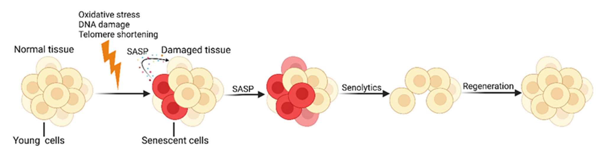
Figure 1. 1. Cellular Cellular senescence and senolytics. SCs continuously produce numerous pro-inflammatory senescence and senolytics. SCs continuously produce numerous pro-inflammamolecules and tissue-remodeling molecules, known as the SASP, which further accelerates the aging tory molecules and tissue-remodeling molecules, known as the SASP, which further accelerates the process. Senolytics promote the regeneration of new healthy cells by identifying and clearing SCs. Created with BioRender.com (accessed on 10 May 2024).

…….omissis

Summary and Prospects
The global issue of population aging is becoming increasingly severe, with elderly individuals being more susceptible to infections and age-related diseases, leading to higher morbidity and mortality rates [5]. Cellular senescence and immunosenescence are closely linked to aging; therefore, this review focuses on immunotherapies targeting aging. It revisits significant recent discoveries in the mechanisms of cellular senescence and immunosenescence that have propelled the development of new treatment paradigms for aging and age-related diseases.
Recent Advances in Aging and Immunosenescence: Mechanisms and Therapeutic Strategies. Shuaiqi Wang1 . Cell. 2025
Department of Immunology, CAMS Key Laboratory T-Cell and Cancer Immunotherapy, Institute of Basic Medical Sciences, Chinese Academy of Medical Sciences and School of Basic Medicine, Peking Union Medical College, State Key Laboratory of Common Mechanism Research for Major Diseases, Beijing 100005, China; b2023005058@pumc.edu.cn (S.W.); huotong1998@pumc.edu.cn (T.H.); b2022005055@student.pumc.edu.cn (M.L.); s2022005053@student.pumc.edu.cn (Y.Z.); zhangjianmin@ibms.pumc.edu.cn (J.Z.). https://doi.org/10.3390/cells14070499

3 – Chronic Low-Grade Inflammation and Brain Structure in the Middle-Aged and Elderly Adults. 2024
Abstract: Low-grade inflammation (LGI) mainly acted as the mediator of the association of obesity and inflammatory diet with numerous chronic diseases, including neuropsychiatric diseases. However, the evidence about the effect of LGI on brain structure is limited but important, especially in the context of accelerating aging. This study was then designed to close the gap, and we leveraged a total of 37,699 participants from the UK Biobank and utilized inflammation score (INFLA-score) to measure LGI. We built the longitudinal relationships of INFLA-score with brain imaging phenotypes using multiple linear regression models. We further analyzed the interactive effects of specific covariates. The results showed high level inflammation reduced the volumes of the subcortex and cortex, especially the globus pallidus ( β [95% confidence interval] = −0.062 [−0.083, −0.041]), thalamus (−0.053 [−0.073, −0.033]), insula (−0.052 [−0.072, −0.032]), superior temporal gyrus (−0.049 [−0.069, −0.028]), lateral orbitofrontal cortex (−0.047 [−0.068, −0.027]), and others. Most significant effects were observed among urban residents. Furthermore, males and individuals with physical frailty were susceptive to the associations. The study provided potential insights into pathological changes during disease progression and might aid in the development of preventive and control targets in an age-friendly city to promote great health and well-being for sustainable development goals.

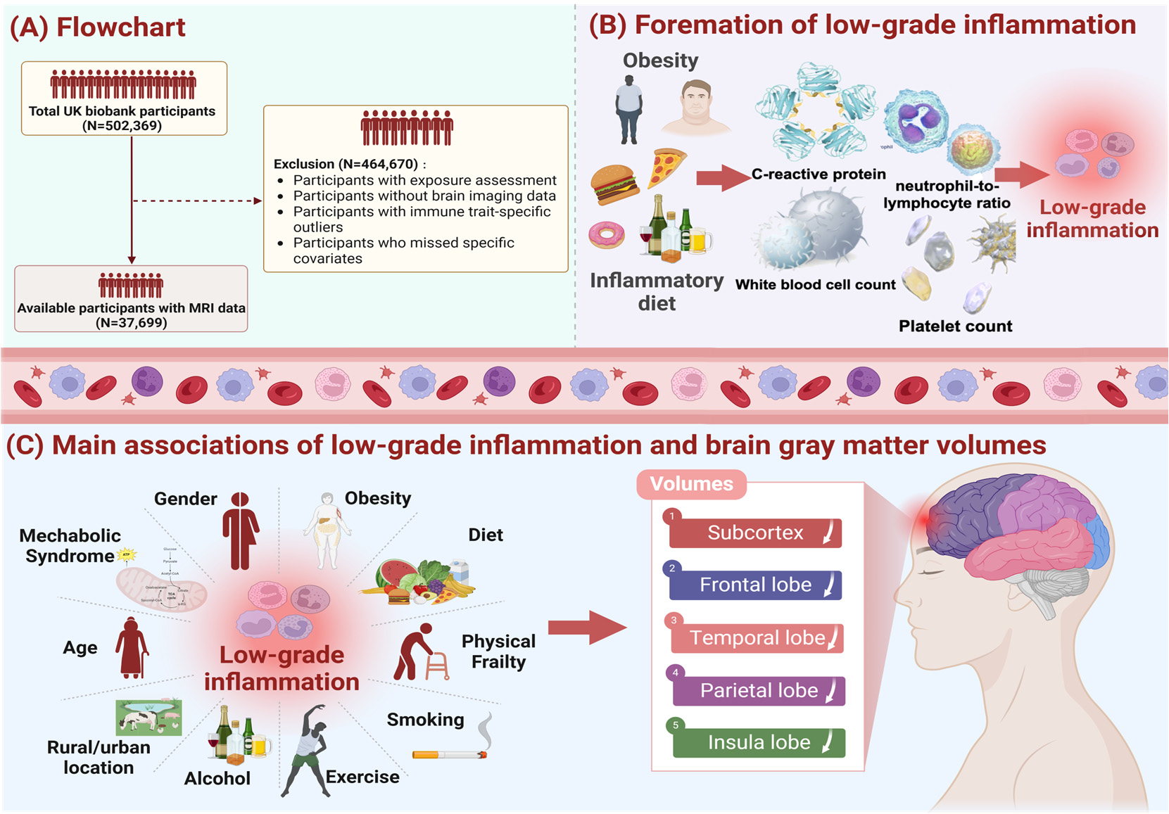
Figure 1. Study workflow.Figure 1. Study workflow. We screened 37,699 UK Biobank participants to explore the effects of
We screened 37,699 UK Biobank participants to explore the effects of low-
grade inflammationlow-grade(LGI) oninflammation (LGI) on thd brain system, and the exclusion criteria of our study population
thd brain system, and the exclusion criteria of our study population is
shown in pane (A).is shownAdditionally,in pane we(A).usedAdditionally,the weINFLA-score,used thecharacterizedINFLA-score, bycharacterizedC-reactivebyprotein,C-reactive protein, white blood cell, plateletwhite bloodcounts,cell,andplateletneutrophil-to-lymphocytecounts, and neutrophil-to-lymphocyteratio, to measureratio,andto measurequantifyandthequantify the levels of LGI, and relevantlevels of LGI,informationand relevantas showninformationin paneas shown(B). Asinshownpane (B).inAspaneshown(C),intakingpane (C),influen-taking influential tial factors of LGI intofactorsaccount,of LGIweintofit theaccount,multiplewe fitlinearthe multipleregressionlinearmodelregressioncontrollingmodel forcontrollingcovariatesfor covariates (age, sex, IMD, WHR,(age,healthysex, IMD,lifestyle,WHR, healthyprevalencelifestyle,of hypertension,prevalence of hypertension, diabetes mellitus and stroke)
diabetes mellitus and stroke)
and conducted subgroupand conductedanalysis bysubgroupage, sex,analysisWHR,bymetabolicage, sex,syndrome,WHR, metabolicphysicalsyndrome,frailty. Thephysicalmainfrailty. The results demonstratedmaina significantresults demonstratedassociationa ofsignificantLGI withassociationatrophyofofLGIbrainwithregions,atrophy ofincludingbrain regions,sub- including cortex, frontal lobe,subcortex,temporalfrontallobe, parietallobe, temporallobe andlobe,insulaparietallobe.lobe and insula lobe.

…omissis

Conclusions
To sum up, the conceptual and design framework of our investigation is to characterize the associations between LGI and brain imaging phenotypes, thus showing that LGI may lead to subclinical cognitive decline or neuropsychic diseases partly via structural neural pathways. Moreover, our analyses revealed that more significant associations of LGI with the atrophy of brain structure among male or individuals with physical frailty. These findings not only contribute to the evolvement of clinical diagnosis and therapy, but also provide a novel perspective for the development of new preventive strategies, namely, when brain lesions are subclinical and without any apparent clinical sign, inflammatory intervention, such as diet therapy, is an early preventive strategy.
Chronic Low-Grade Inflammation and Brain Structure in the Middle-Aged and Elderly Adults. Yujia Bao et al. School of Public Health, Shanghai Jiao Tong University School of Medicine, Shanghai 200025, China; bubble-y@sjtu.edu.cn (Y.B.); c.cctx@sjtu.edu.cn (X.C.); melody321@sjtu.edu.cn (Y.L.); scp-173@sjtu.edu.cn (S.Y.). Nutrients 2024, 16, 2313. https:// doi.org/10.3390/nu16142313

Vitamina D e vitamina C favoriscono un invecchiamento sano

by luciano

Panoramica. Sia la vitamina D che la vitamina C contribuiscono alla salute della barriera intestinale supportando la funzione delle giunzioni strette e promuovendone la riparazione. La vitamina D, attraverso il suo recettore (VDR), regola le proteine che formano le giunzioni strette, che mantengono l’integrità della barriera e modulano il sistema immunitario. La vitamina C può anche promuovere la riparazione della barriera, potenzialmente regolando le vie di segnalazione di Notch e influenzando la composizione del microbioma intestinale, che può ulteriormente supportare la barrier

Ruolo della vitamina D
Giunzioni strette:
La forma attiva della vitamina D, 1,25(OH)2D3, regola l’espressione di proteine ​​come le claudine e la ZO all’interno del complesso delle giunzioni strette, che sono cruciali per il mantenimento e la riparazione della barriera intestinale.
Modulazione immunitaria:
La vitamina D si lega ai recettori VDR nelle cellule immunitarie e modula le risposte immunitarie, contribuendo a proteggere da condizioni che possono compromettere la barriera, come le malattie infiammatorie intestinali.
Integrità della barriera:
Legandosi ai recettori VDR e influenzando le cellule immunitarie e le cellule epiteliali, la segnalazione della vitamina D contribuisce a mantenere una barriera intestinale sana e stabile.

Ruolo della vitamina C
Riparazione della barriera:
L’integrazione di vitamina C ha dimostrato effetti benefici sulla barriera intestinale, contribuendo a riparare i danni.
Segnalazione Notch:
In combinazione con la vitamina D, la vitamina C può regolare la via di segnalazione Notch per proteggere la barriera mucosa intestinale, inclusa l’espressione della claudina-2.
Microbioma intestinale:
L’integrazione di vitamina C può aiutare a bilanciare il microbiota intestinale in individui sani con livelli di vitamina C non ottimali, il che può indirettamente giovare alla barriera riducendo la presenza di batteri produttori di LPS potenzialmente dannosi.

Effetti combinati
Protezione sinergica:
La ricerca indica che la combinazione di vitamina C e vitamina D può offrire maggiori effetti protettivi sulla barriera intestinale rispetto all’assunzione di una delle due vitamine da sola, probabilmente attraverso la loro influenza combinata sulla via di segnalazione Notch.
Potenziale terapeutico:
Entrambe le vitamine sono oggetto di studio per il loro potenziale nella gestione delle patologie intestinali, migliorando l’integrità della barriera e modulando la risposta immunitaria nell’intestino.

Researches

1 – Gut-interplay: key to mitigating immunosenescence and promoting healthy ageing. 2025
Abstract
Background Immunosenescence is the loss and change of immunological organs, as well as innate and adaptive immune dysfunction with ageing, which can lead to increased sensitivity to infections, age-related diseases, and cancer. Emerging evidence highlights the role of gut-vitamin D axis in the regulation of immune ageing, influencing chronic inflammation and systemic health. This review aims to explore the interplay between the gut microbiota and vitamin D in mitigating immunosenescence and preventing against chronic inflammation and age-related diseases.
Main text
Gut microbiota dysbiosis and vitamin D insufficiency accelerate immunosenescence and risk of chronic diseases. Literature data reveal that vitamin D modulates gut microbiota diversity and composition, enhances immune resilience, and reduce systemic inflammation. Conversely, gut microbiota influences vitamin D metabolism to promote the synthesis of active vitamin D metabolites with implications for immune health.
Conclusions
These findings underscore the potential of targeting gut-vitamin D axis to modulate immune responses, delay the immune ageing, and mitigate age-related diseases. Further research is needed to integrate vitamin D supplementation and microbiome modulation into strategies aimed at promoting healthy ageing.
Keywords Gut microbiota, Vitamin D, Immune ageing, Immunosenescence, Healthy ageing
Gut-vitamin D interplay: key to mitigating immunosenescence and promoting healthy ageing. 2025. Hammad Ullah. https://doi.org/10.1186/s12979-025-00514-y
Hammad Ullah hammadrph@gmail.com 1 School of Pharmacy, University of Management and Technology, Lahore 54000, Pakistan

2 – Perspectives About Ascorbic Acid to Treat Inflammatory Bowel Diseases. Ian Richard Lucena Andriolo et al. 2024.

It is known that reactive oxygen species cause abnormal im- mune responses in the gut during inflammatory bowel dis- eases (IBD). Therefore, oxidative stress has been theorized as an agent of IBD development and antioxidant compounds such as vitamin C (L-ascorbic acid) have been studied as a new tool to treat IBD. Therefore, the potential of vitamin C to treat IBD was reviewed here as a critical discussion about this field and guide future research. Indeed, some preclinical studies have shown the beneficial effects of vitamin C in models of ulcerative colitis in mice and clinical and experimental findings have shown that deficiency in this vitamin is associated with the de- velopment of IBD and its worsening. The main mechanisms that may be involved in the activity of ascorbic acid in IBD in- clude its well-established role as an antioxidant, but also others diversified actions. However, some experimental studies em- ployed high doses of vitamin C and most of them did not per- form dose-response curves and neither determined the mini- mum effective dose nor the ED50. Allometric extrapolations were also not made. Also, clinical studies on the subject are still in their infancy. Therefore, it is suggested that the research agenda in this matter covers experimental studies that assess the effective, safe, and translational doses, as well as the ap- propriate administration route and its action mechanism. After that, robust clinical trials to increase knowledge about the role of ascorbic acid deficiency in IBD patients and the effects of their supplementation in these patients can be encouraged.
Perspectives and conclusion
The pathogenesis of IBD is closely related to oxidative stress due to an intense inflammatory insult and the use of vitamin C in IBD, as well as the role of its deficiency, is currently being investigated. Therefore, this perspective reviewed the pharmacological poten- tial of this vitamin to treat and prevent these diseases. In this ap- proach, Vitamin C may help the integrity of the intestinal barrier under the inflammatory stimulus, and enhance intestinal mucosal barrier function, while reducing oxidative stress.
However, a point that is worthy of attention in non-clinical stud- ies presented here is the dose used, which must be adequate for extrapolation in humans. Studies suggest that a daily intake of vi- tamin C from 100 to 400 mg promotes 100 % of the bioavailability and reaches a maximum serum content of 70–80 µmol/L [33, 34]. In addition, when the intake of vitamin C exceeds 500 mg/day, a further increase in plasma concentration is inhibited and when doses greater than 1,000 mg of ascorbic acid are administered in a single dose the bioavailability can decrease by 30 % [34]. This oc- curs because when 500–1,000 mg of vitamin C are administered orally, the intestinal transporter quickly achieves its maximal satu- ration, while the vitamin is progressively excreted by urine [34, 35].
Another important point, which has not yet been studied, is the impact of the pH of the ascorbic acid solution used in the experi- mental studies. Since the pH of an ascorbic acid solution is very low it is expected that its administration can reduce the pH at the in- jection site, intestine, and colon if an enema was used. So, further studies need to address this bias and evaluate the use of buffered ascorbic acid solutions. Perspectives About Ascorbic Acid to Treat Inflammatory Bowel Diseases. Ian Richard Lucena Andriolo et al. 2024. DOI 10.1055/a-2263-1388. ISSN 2194-9379

Permeabilità intestinale, microbiota, dieta ed esercizio fisico II parte

by luciano

Una ricerca recente e approfondita sull’influenza del microbiota intestinale, della dieta e dell’esercizio fisico sulla permeabilità intestinale. Tetiana R. Dmytriv et al. 2024. DOI 10.3389/fphys.2024.1380713.

6 Exercise as a regulator of intestinal barrier integrity

Regular moderate physical exercises are one of the most common recommendations for the prevention of various pathologies, including disruption of the integrity of the intestinal barrier. This may be due to the influence of the gut microbiota. In particular, exercises have been found to increase gut bacterial diversity (Hintikka et al., 2023). However, effects of physical exercises depend on their intensity. For example, endurance athletes have a high incidence of gastrointestinal disorders and the “leaky” gut is one of the most common disorders (Ribeiro et al., 2021). It is characterized by dysfunction of the intestinal epithelial barrier and its excessive permeability. This results in penetration of harmful microorganisms, toxins or undigested food particles into the bloodstream and has a negative effect on health of the whole organism (Aleman et al., 2023).
The effect of exercise on intestinal permeability depends on its duration and intensity. For example, people who exercise frequently and intensely have the same mortality rates as people who lead a sedentary lifestyle (Van Houten et al., 2015). A 60 min bout of intensive treadmill running increased the permeability of the small intestine in runners, whereas low-intensity running had no such effect (Pals et al., 1997). Using the overtraining model with male C57BL/6 mice, it was established that exhaustive exercise exacerbated intestinal inflammation, disrupted integrity and enhanced intestine wall permeability (Hou et al., 2020). Sustained strenuous exercise in racing sled dogs increased the intestinal permeability and the frequency of gastric erosions or ulcerations (Davis et al., 2005). High-intensity interval running increased intestine wall permeability and intestinal-fatty acid binding protein (I-FABP) release in male runners (Pugh et al., 2017). I-FABP is a cytoplasmic protein expressed exclusively in the enterocytes of the small intestine and its increased concentration in the blood is used as a marker of damage to intestinal epithelial cells (Sikora et al., 2019).
Physical exercise of low/moderate intensity can often have positive effects and can be considered as a method of non-pharmacological intervention in inflammatory bowel disease (Ordille and Phadtare, 2023). For example, mice that swam for 30 min before inducing intestinal barrier dysfunction had less intestinal dysfunction compared to mice that had not swum before. This might happen due to a strengthening of antimicrobial function of the intestine as a result of the increase in expression of antimicrobial peptides (Luo et al., 2014). Obese mice that were trained on a motorized treadmill for 45 min per day 5 days a week for 12 weeks had higher expression levels of colonic ZO-1 and occludin. Moderate exercise effectively prevented the development of dysbacteriosis caused by the HFD, as well as intestinal pathology (Wang et al., 2022). Dysbacteriosis and impaired intestinal barrier integrity induced by HFD in wild type mice was prevented by exercise. Exercise on a motor-driven rodent treadmill for 5 days a week for a total of 15 weeks significantly reversed the pathological changes. Ablation of Sestrin 2 protein attenuated the protective effects of exercise, suggesting its involvement in regulation of intestinal permeability (Yu et al., 2022). Thus, it can be concluded that high-intensity exercises often have a negative effect on the integrity of the intestine, whereas low- and moderate-intensity regular exercise can have a positive effects. It may be speculated that moderate damage to the intestinal wall is a hormetic factor that may be used to train organisms to cope with severe damaging challenges. This may be used to increase the adaptive potential of organisms to prevent damaging effects of any stresses of physical and chemical nature on the integrity of the intestinal wall.


6.1 Exercise-induced heat stress

It is known that physical exertion causes heat stress and associated dysfunction of gut integrity. A systematic review examining the relationship between an exercise-induced increase in core body temperature and intestinal permeability demonstrated that the magnitude of exercise-induced hyperthermia correlated with increased intestinal permeability (Pires et al., 2017). An increase in body temperature is a signal to activate the expression of heat shock proteins (HSP) which constitutively function as molecular chaperones maintaining the native structure of the proteins. Their expression is mainly triggered by heat shock signals. During exercise, the level of HSP70 and HSP90 increase (Krüger et al., 2019). Expression of HSP is regulated at the level of heat shock factors (HSF) such as HSF1 that is expressed in all mammalian tissues. Normally it resides in the cytoplasm as a monomer. In response to stressful conditions, it trimerizes, translocates into the nucleus, binds to the heat shock element of target genes and activates the transcription of HSPs, including HSP70/90 (Noble and Shen, 2012).
In this way, exercises cause a homeostatic imbalance, while regular training is adaptive and decreases the degree of this imbalance. Potentially, a higher adaptive steady-state level of HSPs due to regular training could explain their positive effect on gut integrity. At that time, during acute physical exertion, HSPs probably cannot cope with that level of homeostatic imbalance caused exercise-induced heat stress.

6.2 Exercise-induced hypoxia
It is well known, that exercise causes a redistribution of blood flow between tissues. This leads to the development of hypoxia (decreased oxygen levels) in intestinal epithelial cells and activation of hypoxia-inducible factor alpha (HIF-1α) (Wu et al., 2020). Figure 3 schematically shows the influence of exercise-induced hypoxia on intestinal permeability. In normoxia (normal oxygen levels), prolyl hydroxylase hydroxylates HIF-1α at two proline residues (Pro 402 and Pro 564). This results in ubiquitination followed by subsequent proteasomal degradation of HIF-1α (Lee et al., 2004).

…..omissis

7 Conclusion and perspectives
The intestinal wall is a kind of checkpoint between the external and internal environments of organisms. The wall consists of three layers: mucous, epithelial, and lamina propria. The mucous layer is inhabited by microorganisms, many of which mutually beneficially coexistence within the human body. These microorganisms modulate many if not most living processes: from the development of the immune and nervous systems at early stages of life to the induction of chronic inflammation causing neurodegeneration at aging. Despite the fact that these microorganisms have coexisted with humans for many years, under certain conditions the enteral immune system of the lamina propria can perceive them as foreign and trigger a pro- inflammatory response.
Normally, the intestinal mucosa is semipermeable. It allows selective absorption of nutrients into the bloodstream but prevents the entrance of potentially harmful microorganisms and their waste products from contact with the enteral immune system. An imbalance of the intestinal microbiota, called dysbiosis, can cause a disturbance of intestinal integrity and increase intestinal permeability. Conversely, a healthy composition of the gut microbiota can contribute to the integrity of the intestinal barrier due to increased expression and induction of the assembly of TJ proteins, activation of mucus synthesis, and antioxidant action.
Disruption of intestinal barrier function may trigger development of local and even systemic inflammation.
…..omissis
In general, a vicious cycle of intestinal barrier disruption can be traced here, as excessive intestinal wall permeability provokes the development of chronic low-grade inflammation. The latter is characterized by increased production of pro-inflammatory cytokines and enhanced ROS generation, increasing intestinal barrier dysfunction.
Nutrition looks to be the simplest non-pharmacological effector of integrity and permeability of the intestinal wall. It can have both a negative effect, such as HFD inducing metabolic endotoxemia, or a positive effect, such as a diet rich in plant polyphenols or fermented dairy products, increasing the expression of TJ proteins and promoting the development of beneficial bacteria.
Exercise also can affect gut intestinal permeability. Its effects depend on duration and intensity of exercise. Acute extensive physical exertion often increases intestinal permeability which may be related to the induction of heat stress, that organisms cannot cope with at that time due to insufficient resources. On the other hand, regular low and moderate intensity exercises, that are adaptive in nature, mostly have a positive effect on the integrity of the intestine and decrease its permeability. Potentially, this may be associated with an increase in the steady-state level of HSPs and chronic activation of HIF-1α which activates the transcription of genes responsible for strengthening the intestinal barrier function.
In general, it can be concluded that proper nutrition which promotes a healthy biodiversity of the gut microbiota, combined with moderate exercise, contribute to the integrity of the intestine. Disbalanced nutrition and excessive physical activity can provoke the development of dysbacteriosis and increase intestinal permeability which can potentially lead to a pro-inflammatory response. Figure 4 schematically shows potential consequences of acute intense exercises, unhealthy diet (e.g., high-fat diet), and dysbiosis on the intestinal barrier.
Taking into account all of the above, we can outline the following future prospects:
1. Development of healthy diets to support intestinal homeostasis;
2. Use of fermented dairy products as natural pre-, pro- and postbiotics to promote a healthy gut;
3. Selection of exercises to promote intestinal integrity by frequency, intensity and duration;
4. Study of the role of intestinal HIF-2α during exercise;
5. Systemic investigation of hypoxia-induced oxidative stress as a regulator of intestinal wall permeability.
Most of these perspective avenues are directed to enhance the capability of organisms to cope with disturbing factors. That increases an adaptive capability via preadaptation/hormetic mechanisms. However, some of them may be used “to patch holes” in “leaky” intestinal wall, which is characterized by increased specific permeability of the intestinal epithelium. Intestinal barrier permeability: the infuence of gut microbiota, nutrition, and exercise. Tetiana R. Dmytriv et al. DOI 10.3389/fphys.2024.1380713. PUBLISHED 08 July 2024

Note
[1] The term “intestinal barrier” emphasizes the barrier function of the intestinal wall which protects organism against invading by bacteria or other microorganisms and potentially toxic components of microorganisms. In fact, it is a complex selective physical barrier that separates the internal environment of the body from the contents of the intestinal lumen (Bischoff et al., 2014). Figure 1 shows a schematic structure of the intestinal barrier. It consists of several layers: i) a mucous layer including inner and outer mucous sublayers inhabited by commensal microorganisms in a different extent, ii) a single layer of epithelial cells, and iii) the lamina propria, which consists of immune cells that instantly react to the invasion of foreign substances (Schoultz and Keita, 2020).
The first layer, the mucous layer, that consists mainly of a mesh polymer called mucin, is located on the side of the intestinal lumen. It is associated with community of commensal microorganisms, including bacteria, fungi, viruses, and parasites, that form the individual microbial community (Chelakkot et al., 2018). A change in the microbial composition that causes a sharp imbalance between beneficial and potentially pathogenic bacteria, including changes in its functional composition, metabolic activity or changes in their local distribution, is called dysbiosis or dysbacteriosis. The latter usually results from loss of beneficial bacteria, overgrowth of potentially pathogenic bacteria, or loss of overall bacterial diversity. This disrupts the homeostatic balance of the intestinal microbiota and has a negative impact on the host’s health. In particular, dysbacteriosis is implicated in a wide range of diseases (DeGruttola et al., 2016).
The second layer, the intestinal epithelium, consists of a single layer of several specialized epithelial cells, such as enterocytes, Goblet cells, Paneth cells, enteroendocrine cells, and microfold cells (Figure 1). Enterocytes form the basis of the intestinal epithelium and play a main role in the absorption of all consumed nutrients. Goblet cells constitute about 10% of specialized epithelial cells. They secrete mucus to protect the intestinal wall from digestive enzymes (Kim and Ho, 2010). Paneth cells contain secretory granules filled with antimicrobial peptides, that are secreted in low amounts constitutively and provide the antimicrobial properties of the intestinal mucosa. Under certain conditions, their secretion can increase dramatically (Yokoi et al., 2019). Enteroendocrine cells produce hormones regulating secretion of digestive enzymes and insulin, peristalsis of the intestine, satiety, and immune response (Bonis et al., 2021). Microfold cells transport bacteria and antigens from the epithelium to enteric immune cells that either activate or suppress the immune response (Jung et al., 2010). All these cell types collectively contribute significantly to gut homeostasis.
The third layer, lamina propria, is located under the epithelium and forms the enteric immune system that consists of a large number of leukocytes with macrophages and dendritic cells being the dominant cell types (Shemtov et al., 2023). Resident intestinal macrophages are located in close proximity to the gut microbiota, with which they often interact. They play a key role in immune sampling of luminal bacteria, contributing to the maintenance of intestinal homeostasis and regulated immune response.

[2] TJ proteins are a complex of transmembrane and cytoplasmic proteins that form tight junctions, which seal cells together to create a selective barrier, maintain cell polarity, and regulate cell processes.

Key words
tight junction, tight junction proteins, inflammation,

Permeabilità intestinale, microbiota, dieta ed esercizio fisico I parte

by luciano

Una ricerca recente e approfondita sull’influenza del microbiota intestinale, della dieta e dell’esercizio fisico sulla permeabilità intestinale. Tetiana R. Dmytriv et al. 2024. DOI 10.3389/fphys.2024.1380713.

In Evidenza
1. La parete intestinale [1] è composta da tre strati: mucosa, epiteliale e lamina propria. Lo strato mucoso è abitato da microrganismi, molti dei quali coesistono reciprocamente beneficamente all’interno del corpo umano. Questi microrganismi modulano molti se non la maggior parte dei processi viventi: dallo sviluppo del sistema immunitario e nervoso nelle prime fasi della vita all’induzione dell’infiammazione cronica che causa neurodegenerazione nell’invecchiamento. Nonostante il fatto che questi microrganismi abbiano coesistito con gli esseri umani per molti anni, in determinate condizioni il sistema immunitario enterale della lamina propria può percepirli come estranei e innescare una risposta pro-infiammatoria.

2. Normalmente, la mucosa intestinale è semipermeabile. Consente l’assorbimento selettivo dei nutrienti nel flusso sanguigno, ma impedisce l’ingresso di microrganismi potenzialmente dannosi e dei loro prodotti di scarto dal contatto con il sistema immunitario enterale. Uno squilibrio del microbiota intestinale, chiamato disbiosi, può causare un disturbo dell’integrità intestinale e aumentare la permeabilità intestinale.

3. L’eccessiva permeabilità della parete intestinale provoca lo sviluppo di un’infiammazione cronica di basso grado.

4. La nutrizione sembra essere il più semplice agente non farmacologico di integrità e permeabilità della parete intestinale. Può avere sia un effetto negativo, come l’HFD che induce l’endotossemia metabolica, sia un effetto positivo, come una dieta ricca di polifenoli vegetali o prodotti lattiero-caseari fermentati, aumentando l’espressione delle proteine TJ [2] e promuovendo lo sviluppo di batteri benefici.

5. L’esercizio fisico può anche influenzare la permeabilità intestinale. I suoi effetti dipendono dalla durata e dall’intensità dell’esercizio. Lo sforzo fisico acuto esteso spesso aumenta la permeabilità intestinale che può essere correlata all’induzione dello stress da calore, che gli organismi non possono far fronte in quel momento a causa delle risorse insufficienti. D’altra parte, gli esercizi regolari di bassa e moderata intensità, che sono di natura adattiva, hanno per lo più un effetto positivo sull’integrità dell’intestino e ne riducono la permeabilità.

La ricerca
“La parete intestinale è una barriera selettivamente permeabile tra il contenuto del lume intestinale e l’ambiente interno del corpo. I disturbi della permeabilità della parete intestinale possono potenzialmente portare a un’attivazione indesiderata del sistema immunitario enterico a causa di un contatto eccessivo con il microbiota intestinale e i suoi componenti e lo sviluppo di endotossemia, quando il livello di lipopolisaccaridi batterici aumenta nel sangue, causando infiammazione cronica a bassa intensità. In questa revisione, vengono trattati i seguenti aspetti: la struttura della barriera della parete intestinale; l’influenza del microbiota intestinale sulla permeabilità della parete intestinale attraverso la regolazione del funzionamento delle proteine a giunzione stretta, la sintesi/degradazione del muco e degli effetti antiossidanti; i meccanismi molecolari di attivazione della risposta proinfiammatoria causata dall’invasione batterica attraverso le cascate di segnalazione TIRAP/MyD88 e TRAM/TRIF indotte da TLR4; l’influenza della nutrizione sulla permeabilità intestinale e l’influenza dell’esercizio fisico con un’enfasi sullo stress da calore indotti dall’esercizio e sull’ipossia. Nel complesso, questa revisione fornisce alcune informazioni su come prevenire l’eccessiva permeabilità della barriera intestinale e i processi infiammatori associati coinvolti in molte, se non nella maggior parte delle patologie. Alcune diete e l’esercizio fisico dovrebbero essere approcci non farmacologici per mantenere l’integrità della funzione di barriera intestinale e fornire il suo funzionamento efficiente. Tuttavia, in tenera età, l’aumento della permeabilità intestinale ha un effetto ormetico e contribuisce allo sviluppo del sistema immunitario.

Introduzione

La parete intestinale è un sistema complesso composto da quattro strati: mucosa, sottomucosa, muscolo e serosa. Il termine “barriere intestinale” enfatizza la componente protettiva della parete intestinale, mentre la permeabilità intestinale è una caratteristica misurabile dello stato funzionale della barriera intestinale (Bischoff et al., 2014). La parete fornisce un assorbimento selettivo di nutrienti e altri componenti del lume intestinale. Allo stesso tempo, la barriera intestinale protegge il corpo dall’ingresso di sostanze estranee indesiderate, particelle di cibo, microrganismi e loro componenti. Negli organismi normalmente funzionanti, la permeabilità della parete intestinale è strettamente controllata, ma il suo disturbo, se non adeguatamente fissato, può portare a molte, se non la maggior parte, patologie acquisite (Gieryńska et al., 2022).

Il tratto gastrointestinale (GIT) è abitato da diversi microbi chiamati microbiota intestinale che formano una comunità molto dinamica.

Figura 1 La struttura schematica della barriera intestinale. Per i dettagli vedi il testo.

L'”Ipotesi dei vecchi amici” suggerisce che le persone si sono evolute con molti microbi che, oltre a molte funzioni fisiologiche, stimolano anche lo sviluppo del sistema immunitario e ne regolano il funzionamento (Rook, 2023). Gli antigeni microbici sono sotto costante sorveglianza da parte del sistema immunitario enterico. Le cellule T immunitarie regolatorie sono responsabili del mantenimento della tolleranza immunitaria del microbiota intestinale omeostatico (Wu e Wu, 2012). Tuttavia, l’aumento della permeabilità intestinale può promuovere la traslocazione dei batteri luminali e dei modelli molecolari associati ai microbi, in particolare i lipopolisaccaridi (LPS) dall’intestino al flusso sanguigno, innescando lo sviluppo di endotossemia e infiammazione cronica a bassa intensità (Vanuytsel et al., 2021). L’endotossemia indotta dalla dieta è definita come endotossemia metabolica. Ad esempio, Cani et al. (2007) hanno stabilito che una dieta ricca di grassi ha aumentato cronicamente le concentrazioni plasmatiche di LPS da due a tre volte.

I lipopolisaccaridi endogeni LPS vengono costantemente rilasciati a causa della morte di batteri Gram-negativi nell’intestino. Con un aumento della permeabilità della barriera intestinale, gli LPS vengono assorbiti nel flusso sanguigno portale, da dove vengono trasportati dalle lipoproteine direttamente nel fegato, formando l’asse intestino-fegato. Inoltre, sono metabolizzati dagli enzimi epatici ed escreti con la bile. Tuttavia, se la loro degradazione o l’escrezione biliare sono compromesse, l’LPS può raggiungere la circolazione sistemica, dove si lega al recettore Toll-like 4 (TLR4) su leucociti, cellule endoteliali e piastrine, causando infiammazione arteriosa. In definitiva, questo porta all’attivazione della coagulazione del sangue e alla formazione di trombi, che dimostra che l’infiammazione indotta da LPS associata all’aumento della permeabilità della parete intestinale può essere coinvolta nello sviluppo dell’aterosclerosi e delle malattie trombotiche (Violi et al., 2023). In generale, la rottura della funzione di barriera intestinale è coinvolta in molte malattie correlate e non correlate al GIT, tra cui la malattia infiammatoria intestinale, la malattia epatica associata alla disfunzione metabolica, il malassorbimento degli acidi biliari, la celia, il diabete di tipo I, l’obesità, la schizofrenia e altre (Vanuytsel et al., 2021). Potenzialmente, questo potrebbe essere superato da un intervento non farmacologico basato su dieta ed esercizi (Pražnikar et al., 2020; Ordille e Phadtare, 2023) che promuovono un ecosistema intestinale sano e alleviano i sintomi di molte patologie.

In questa recensione, descriviamo la struttura della parete intestinale e i meccanismi molecolari della risposta pro-infiammatoria causata dall’invasione batterica a causa del disturbo della permeabilità della parete intestinale, nonché le influenze del microbiota intestinale, della dieta e degli esercizi sulla permeabilità della parete intestinale. Diete specifiche ed esercizi regolari a bassa e moderata intensità sono proposti come approcci non farmacologici efficaci per mantenere l’integrità della parete intestinale e il suo funzionamento efficiente. Tuttavia, in tenera età, la perdita controllata dell’intestino può essere necessaria per innescare lo sviluppo del sistema immunitario attraverso meccanismi ormetici.

2 La struttura della barriera intestinale

…….omissis

3 Permeabilità intestinale

La semipermeabilità o permeabilità selettiva è una caratteristica cruciale della parete intestinale. Limita la penetrazione degli agenti patogeni ma consente la permeabilità di nutrienti, acqua e ioni. I fattori endogeni (ad esempio, infiammazione) ed esogeni (ad esempio, componenti dietetici, sostanze tossiche o farmaci) possono aumentare la permeabilità intestinale e causare la formazione di un cosiddetto “intestino permeabile”. Quest’ultimo è caratterizzato dalla penetrazione di antigeni alimentari, commensali o batteri patogeni nel sangue, causando lo sviluppo di infiammazione (Vanuytsel et al., 2021). Alcune malattie possono anche agire come fattore dirompente della barriera intestinale. Ad esempio, diversi studi dimostrano che l’iperglicemia, una caratteristica chiave del diabete, induce la disfunzione della barriera intestinale (Thaiss et al., 2018; Dubois et al., 2023). L’esposizione prolungata al glucosio ad alti livelli aumenta la capacità di migrazione della linea cellulare del colon umano Caco-2, con il risultato che gli strati appaiono meno organizzati rispetto alle condizioni fisiologiche. In particolare, questo è associato a una diminuzione dell’espressione delle proteine della giunzione stretta (TJ), che contribuisce all’interruzione della rete strutturale ad esse associata e a un aumento della permeabilità della barriera intestinale (Dubois et al., 2023). A sua volta, questo contribuisce alla penetrazione dei batteri luminali e allo sviluppo della disbatteriosi con conseguente infiammazione. Ad esempio, Harbison et al. (2019) hanno dimostrato che i bambini con diabete di tipo I hanno disbiosi del microbiota intestinale associata ad un aumento della permeabilità intestinale. In particolare, sono stati osservati una minore diversità microbica, un numero inferiore di specie batteriche antinfiammatorie e batteri produttori di SCFA, e questi cambiamenti non sono stati spiegati dalle differenze nella dieta. Pertanto, alcune malattie, tra cui il diabete, possono anche svolgere il ruolo di disgregatori della barriera intestinale.

Mucus and epithelium are the most important components of the intestinal barrier that limit the development of inflammation. The mucous layer consists of two sublayers (Figure 1). The outerlayer is thick and loose. It is inhabited by a large number of commensal microorganisms that form colonies, and under healthy conditions pathogenic bacteria cannot outgrow them or penetrate further. In other words, homeostatic microorganisms efficiently compete with potentially pathogenic ones and prevent their excessive proliferation. The inner sublayer, on the contrary, is solid and contains only a few microbes (Usuda et al., 2021). The gut microbiota plays a major role in changing the composition of mucus, regulating its synthesis and degradation.
Epithelial cells are connected by TJ proteins (Lee et al., 2018) which regulate the absorption of water, ions, and dissolved substances. They include two functional categories of proteins: integral transmembrane proteins, located at the border of adjacent cell membranes, and adaptive peripheral membrane proteins that connect integral proteins with the actin cytoskeleton. The former includes occludin, claudins, junctional adhesion molecules, and tricellulin whereas the latter include zonula occludens-1 (ZO-1), ZO-2, and ZO-3 (Lee et al., 2018). The gut microbiota can influence the expression and localization of all of these TJ proteins.

Risposta del sistema immunitario all’infiammazione di basso grado: relazione con le funzioni metaboliche

by luciano

(Immunology of chronic low-grade inflammation: relationship with metabolic function)
L’infiammazione fa parte della risposta immunitaria innata dell’organismo ed è un processo essenziale che non solo difende da batteri e patogeni nocivi, ma svolge anche un ruolo chiave nel mantenimento e nella riparazione dei tessuti. In condizioni patologiche, si verifica un’interazione bilaterale tra la regolazione immunitaria e il metabolismo aberrante, con conseguente infiammazione persistente in assenza di infezione. Questo fenomeno è definito infiammazione metabolica sterile (metainfiammazione) e si verifica se lo stimolo iniziale non viene rimosso o se il processo di risoluzione viene interrotto.

…..omissis

Questa infiammazione metabolica cronica di basso grado non deve essere trascurata poiché è significativamente associata alla mortalità per tutte le cause nella popolazione generale (Fest et al. 2019), ha un impatto negativo sulla sensibilità all’insulina (Blaszczak et al. 2020) e aumenta il rischio di sviluppo del cancro (Li et al. 2023).
Immunology of chronic low-grade inflammation: relationship with metabolic function. Mari van de Vyver. Division of Clinical Pharmacology, Department of Medicine, Faculty of Medicine and Health Sciences, Stellenbosch University, Cape Town, South Africa. Journal of Endocrinology (2023) 257, e220271

Note:
1 – Funzione metabolica:
La funzione metabolica si riferisce ai processi chimici continui all’interno delle cellule e degli organismi che convertono il cibo in energia utilizzabile, costruiscono e riparano i tessuti e sostengono la vita. Ciò include processi vitali come la respirazione, la circolazione sanguigna e il mantenimento cellulare, anche a riposo. Le due principali categorie di reazioni metaboliche sono l’anabolismo, che costruisce molecole più grandi e consuma energia, e il catabolismo, che scompone molecole più grandi per rilasciare energia, come la digestione del cibo.

2 – L’immunologia dell’infiammazione descrive la risposta del sistema immunitario ai danni tissutali o alle infezioni, un meccanismo di difesa innato e non specifico che serve a eliminare gli agenti nocivi e a promuovere la riparazione. La flogosi, o infiammazione, coinvolge cellule e molecole immunitarie che richiamano più sangue nel sito danneggiato, causando arrossamento, calore, gonfiore e dolore. Sebbene sia un processo protettivo, un’infiammazione eccessiva o cronica può diventare dannosa e contribuire a malattie autoimmuni o altre patologie.