Header Image - Gluten Light

Yearly Archives

13 Articles

Sensibilità al glutine non allergica non celiaca: sintomi

by luciano

La Non-Celiac Gluten Sensitivity (NCGS), aneddoticamente descritta in passato e dal 2010 riconosciuta come una nuova entità clinica, si riferisce a quei pazienti che, senza essere affetti da celiachia né da allergia al frumento IgE mediata [1], presentano una serie di manifestazioni cliniche intestinali ed extraintestinali, che insorgono tempestivamente dopo ingestione di alimenti contenenti glutine e altrettanto rapidamente scompaiono a dieta aglutinata. Nonostante la consapevolezza del dato clinico è una condizione a patogenesi ignota (si ipotizza un ruolo dell’immunità innata) e in cui, in assenza di markers genetici, sierologici e istologi, la diagnosi è largamente ipotetica, posta su base clinica e in base a criteri di esclusione Nei pochi studi presenti in letteratura condotti in cieco vs placebo per verificare la reale risposta all’ingestione di glutine, possono essere sollevate obiezioni metodologiche, i risultati sono controversi, è emerso il ruolo dell’effetto nocebo, e l’effetto glutine- ‐ specifico sembra molto limitato. Da segnalare inoltre recenti evidenze riguardanti molecole, spesso presenti negli stessi alimenti contenenti glutine e in grado di scatenare disturbi sovrapponibili e in comune con la s. dell’intestino irritabile (FODMAPs, fermentable, oligo- ‐, di- ‐ and mono- ‐saccharides and polyols; ATIs, amylase trypsin inhibitors).
Nella NCGS l’esclusione del glutine dalla dieta risolve la sintomatologia entro pochi giorni; nei pazienti che lamentano disturbi dopo ingestione di alimenti contenenti frumento è pertanto necessario escludere sia la celiachia che l’allergia al grano, e quindi confermare la diagnosi attraverso il monitoraggio clinico dopo introduzione di una dieta di esclusione seguita dalla reintroduzione della dieta libera; in assenza di markers specifici e per la verosimile influenza di effetto placebo/nocebo, il percorso diagnostico dovrebbe essere condotto presso le strutture e dalle stesse figure professionali dei Centri di Riferimento e Presidi di Rete per la MC (gastroenterologo dell’adulto o pediatrico, allergo immunologo, specialisti del settore nutrizione).
La prevalenza varia in letteratura dallo 0.6% al 6%, nel 50% dei casi si rileva associazione con gli aplotipi HLA DQ2/DQ8, valore statisticamente non significativo rispetto alla popolazione generale, esiste una netta prevalenza nel sesso femminile e in una variabile percentuale dei pazienti è rilevabile una positività sierologica per gli anticorpi anti gliadina (AGA) di prima generazione, non più utilizzabili per la diagnosi di celiachia per la scarsa accuratezza diagnostica. L’esame istologico della mucosa intestinale risulta nella norma o documenta un aumento dei linfociti intraepiteliali in assenza di atrofia villosa. Sono invece descritti segni di attivazione dell’immunità innata, non glutine- ‐specifica (Tab 4.1).

Quando sospettarla
Nell’età adulta i sintomi possono essere gastrointestinali, assimilabili alla sindrome dell’intestino irritabile oppure reflusso gastroesofageo, nausea, stomatite aftosa [2], epigastralgia [3] (Fig 4.1), associati o meno a sintomi extraintestinali, tra cui prevalgono l’astenia, la confusione mentale, le artralgie e le mialgie, la cefalea, le eruzioni cutanee (Fig.4.2).

Figura 4.1 Sintomi gastrointestinali nella sospetta non-celiac gluten sensitivity (%= percentuale di pazienti)
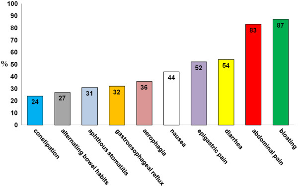
Sintomi gastrointestinali nella sospetta non-celiac gluten sensitivity

Figura 4.2 Sintomi non gastrointestinali nella sospetta non- ‐celiac gluten sensitivity (%= percentuale di pazienti)
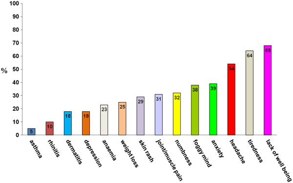
Sintomi non gastrointestinali nella sospetta non- ‐celiac gluten sensitivity

La diagnosi
Al momento la diagnosi di sensibilità al glutine è solo d’esclusione, non esiste un bio-marker specifico né test dedicati, occorre sospettarla quando è possibile dimostrare che la sintomatologia riferita dal paziente è completamente risolta dall’esclusione del glutine e solo del glutine dalla dieta, mentre la sua reintroduzione determina in tempi brevi, ore o giorni, il ripresentarsi dell’intera sintomatologia.
A fronte di questo occorre escludere la MC e l’allergia al grano come già sottolineato.
Nella GS poi non è nota la dose tollerata di glutine né per quanto tempo occorre escludere il glutine dalla dieta: gli studi clinici relativi a questa condizione dovrebbero prevedere l’effettuazione, nelle strutture allergo- ‐immunologiche dedicate, del DBPCT con il glutine; questo test può consentire anche in questa condizione come nell’allergia IgE mediata, un maggior conforto diagnostico e può anche consentire di stabilire quale dose minima può essere tollerata dal singolo individuo.

Note

[1] Allergia alimentare a grano IgE-mediata. I soggetti allergici sviluppano sintomi da minuti a 2 ore dopo l’assunzione di grano. I sintomi includono orticaria, angioe- dema, eritema, prurito, vomito, dolore addominale, tosse, raucedine, wheezing, stridore, distress respiratorio, congestione nasale fino all’anafilassi.
L’allergia IgE-mediata può essere considerata come un’alterazione della risposta immunitaria, ovvero una reazione anormale e specifica verso sostanze, in realtà innocue, percepite come nocive, quindi, attaccate dalle difese immunitarie dell’organismo.
La classe di anticorpi che entra in gioco prende il nome di immunoglobuline E (IgE).
La reazione insorge in seguito a contatto, ingestione o inalazione di sostanze che possono essere di varia natura e che prendono genericamente il nome di allergeni.
La prima esposizione all’allergene determina una sensibilizzazione dell’organismo che produce specifiche immunoglobuline (IgE), senza la comparsa di sintomi. A partire dal secondo contatto, si innescano reazioni a cascata in cui intervengono altri componenti del sistema immunitario, quali i mastociti (a livello tissutale) ed i basofili (a livello ematico).
I mastociti si trovano sotto la superficie cutanea e nelle membrane che rivestono il naso, l’apparato respiratorio, gli occhi e l’intestino. Le IgE, attivate dal legame con l’antigene, vanno a scatenare la risposta allergica legandosi a queste cellule. La degranulazione dei mastociti provoca la liberazione di istamina, leucotrieni e prostaglandine: tali mediatori chimici, agendo su diversi organi e tessuti, provocano l’insorgenza dei sintomi che caratterizzano le risposte allergiche.
È possibile, dunque, identificare tre punti cardine per questa tipologia di reazione:
1. si prevede il coinvolgimento delle IgE;
2. le risposte avvengono rapidamente in seguito al contatto con l’allergene, facilitando il riconoscimento del nesso causa-effetto;
3. lo scatenarsi della risposta è indipendente dalla quantità di allergene con cui l’organismo viene a contatto: si tratta di reazioni NON dose-dipendenti.

[2] (La stomatite aftosa è la comune malattia della bocca, tipica dei giovani e dei giovani adulti, che comporta la ripetuta comparsa di ulcere benigne sulla mucosa orale.)

[3] L’epigastralgia (o dolore alla bocca dello stomaco) è un disturbo molto comune, che si manifesta come un dolore acuto localizzato nella parte superiore dell’addome. Le cause sono molte e legate principalmente a patologie del sistema digestivo.

Bibliografia
• Sapone A, et al. Spectrum of gluten- ‐related disorders: consensus on new
nomenclature and classification. BMC Med 2012;10:13.
• Catassi C, et al. Non- ‐Celiac Gluten sensitivity: the new frontier of gluten
related disorders. Nutrients 2013;5:3839–3853
• Volta U et al. An Italian prospective multicenter survey on patients suspected
of having non- ‐celiac gluten sensitivity. BMC Med. (2014)

Approfondomento:

Esclusione del glutine per i pazienti con NCGS:
Hansen et al. hanno dimostrato che quantità minime di glutine sono sufficienti per influenzare la popolazione del microbiota, abbassando la quantità dei bifidobatteri (batteri buoni) nei pazienti che aderiscono a un regime a basso contenuto di glutine. Leggi di più…..

Dieta senza glutine: effetti

by luciano

In evidenza
1. Omissis…La GFD ha comportato una riduzione delle popolazioni batteriche generalmente considerate benefiche per la salute umana, come Bifidobacterium e Lactobacillus, e un aumento di quelle di patogeni opportunisti come Escherichia coli e Enterobacteriaceae totali.
2. Omissis…Hansen et al. hanno dimostrato che quantità minime di glutine sono sufficienti per influenzare la popolazione del microbiota, abbassando la conta dei bifidobatteri nei pazienti che aderiscono a un regime a basso contenuto di glutine.
3. Omissis…Alcuni cambiamenti nell’abbondanza di 8 famiglie di batteri sono stati osservati durante il periodo della GFD: Veillonellaceae, Ruminococcus bromii e Roseburia faecis, sono diminuiti, mentre Victivallaceae, Clostridiaceae, ML615J-28, Slackia e Coriobacteriaceae sono aumentati durante la GFD. Le Veillonellaceae, una famiglia proinfiammatoria di batteri Gram-negativi noti per la fermentazione del lattato, aumento di malattie come IBD, sindrome dell’intestino irritabile e cirrosi epatica.
4. Omissis….Questa revisione ha valutato le attuali conoscenze sul microbiota intestinale in soggetti sani, nonché su CD e NCG/WS e i relativi effetti provocati dalla dieta senza glutine in queste due condizioni più comuni. Le prove finora acquisite hanno dimostrato che le malattie sono spesso caratterizzate da uno squilibrio nella composizione della popolazione microbica intestinale, che porta alla disbiosi, una condizione che promuove l’infiammazione e il deterioramento metabolico.

Ricerche esaminate:

1 – Effetti di una dieta priva di glutine sul microbiota intestinale e sulla funzione immunitaria in soggetti umani adulti sani. Pubblicato online dalla Cambridge University Press: 18 maggio 2009. Giada De Palma, Inmaculada Nadal, Maria Carmen Collado e Yolanda Sanz
…omissis. “Therefore, introduction of a GFD implied a reduction in bacterial populations generally regarded as beneficial for human health such as Bifidobacterium and Lactobacillus, and an increase in those of opportunistic pathogens such as Escherichia coli and total Enterobacteriaceae. These changes could be related to reductions in polysaccharide intake, since these dietary compounds usually reach the distal part of the colon partially undigested, and constitute one of the main energy sources for beneficial components of the gut microbiota(Reference De Graaf and Venema27). In addition, reductions in Bifidobacterium and Lactobacillus populations relative to Gram-negative bacteria (Bacteroides and Escherichia coli) were previously detected in untreated CD children and particularly in treated CD patients with a GFD(Reference Nadal, Donat and Ribes-Koninckx7). These findings indicate that this dietary therapy may contribute to reduce beneficial bacterial group counts and increase enterobacterial counts, which are microbial features associated with the active phase of CD(Reference Nadal, Donat and Ribes-Koninckx7, Reference Collado, Donat and Ribes-Koninckx28) and, therefore, it would not favour completely the normalisation of the gut ecosystem in treated CD patients”.

2 – Effect of Gluten-Free Diet on Gut Microbiota Composition in Patients with Celiac Disease and Non-Celiac Gluten/Wheat Sensitivity. Giacomo Caio, Lisa Lungaro, Nicola Segata, Matteo Guarino, Giorgio Zoli, Umberto Volta, and Roberto De Giorgio. Nutrients. 2020 Jun; 12(6): 1832. Published online 2020 Jun 19. doi:10.3390/nu12061832
“Celiac disease (CD) and non-celiac gluten/wheat sensitivity (NCG/WS) are the two most frequent conditions belonging to gluten-related disorders (GRDs). Both these diseases are triggered and worsened by gluten proteins ingestion, although other components, such as amylase/trypsin inhibitors (ATI) and fermentable oligosaccharides, disaccharides, monosaccharides and polyols (FODMAPs), seem to be involved in the NCG/WS onset. Therefore, the only effective treatment to date is the long-life adherence to a strictly gluten-free diet. Recently, increasing attention has been paid to the intestinal barrier, a dynamic system comprising various components, which regulate the delicate crosstalk between metabolic, motor, neuroendocrine and immunological functions. Among the elements characterizing the intestinal barrier, the microbiota plays a key role, modulating the gut integrity maintenance, the immune response and the inflammation process, linked to the CD and NCG/WS outbreak. This narrative review addresses the most recent findings on the gut microbiota modulation induced by the gluten-free diet (GFD) in healthy, CD and NCG/WS patients”.
Omissis…..7. Gluten-Free Diet Effects on Healthy Human Microbiota.
The overall literature search on databases including the terms “gluten free diet”, “GFD”, “gluten free diet AND healthy”, “microbiota”, “microbiome”, “microbiome AND healthy patients”, “microbiota AND healthy patients” produced 2775 results. Of these, excluding duplicates, three fulfilled our inclusion criteria. In 2009, De Palma et al. [101] explored whether a month of GFD affects the microbiota composition of ten healthy subjects. Enumeration of fecal bacteria by fluorescence in situ hybridization (FISH) using 16S rRNA-targeted oligonucleotide probes showed that GFD causes a decrease in the count of Bifidobacterium, Clostridium lituseburense and Faecalibacterium prausnitzii. Quantitative PCR (qPCR) characterization of fecal microbes following GFD revealed a reduction in the number of Bifidobacterium, Lactobacillus and Bifidobacterium longum and an increase in the Enterobacteriaceae and Escherichia coli counts. They propose that the depletion in Bifidobacterium and Lactobacillus, generally considered as probiotics, could be caused by the reduced availability of polysaccharides introduced with the GFD that serve as a substrate for gut microbiota. Moreover, the reduction in Faecalibacterium prausnitzii, along with the concomitant increase in the opportunistic pathogens Enterobacteriaceae and Escherichia coli in the fecal mucus of active Crohn’s disease patients was found to trigger the inflammatory insult [89,102,103]. Moreover, Hansen et al. showed that minimal amounts of gluten are sufficient to affect the microbiota population, lowering the Bifidobacteria count in patients adhering to a low-gluten regimen [104]. Indeed, the authors performed a randomized, controlled, cross-over trial study involving 60 non-CD Danish adults who followed a low-gluten diet (2 g gluten per day) for eight weeks and then switched to a high-gluten diet (18 g gluten per day) for another eight weeks, including a washout period of at least six weeks of normal diet (12 g gluten per day) between the two diets. Notably, GFD was associated with an increase of unclassified species of Clostridiales and an unclassified species of Lachnospiraceae, whereas E. hallii and A. hadrus (both butyrate-producers), Dorea (hydrogen producer) and the hydrogen-consumer and acetate-producer Blautia, in addition to two species of the Lachnospiraceae and four species of Bifidobacterium, were found to decrease. These microbial changes could be ascribed to the low-gluten diet availability of arabinoxylan and arabinoxylan-oligosaccharides, as these food components are abundant non-starch polysaccharides of cereal grains, which serve as energy substrates for the bacterial species mentioned above [105,106,107,108,109,110]. Bonder et al. [111] investigated the gut microbiota of 21 healthy volunteers on a GFD for four weeks, tested with a total of 9 stool samples for each person (one at baseline, four during the GFD and four when they returned to their usual diet). The microbiome profile was then characterized using 16 sRNA sequencing and investigated for taxonomic and implied functional compositions. Overall, the bacterial profile remained relatively stable in healthy individuals on GFD. However, some changes in the abundance of 8 families of bacteria were observed during the GFD period: Veillonellaceae, Ruminococcus bromii and Roseburia faecis, decreased, whereas Victivallaceae, Clostridiaceae, ML615J-28, Slackia and Coriobacteriaceae increased during GFD. Veillonellaceae, a pro-inflammatory family of Gram-negative bacteria known for lactate fermentation, increase in diseases such as IBD, irritable bowel syndrome and liver cirrhosis [88,112,113], while they decrease in autistic patients [114]. Compared to a normal diet, the abundance of Ruminococcus bromii, known to degrade the resistant starch in the human colon [115] and the cellulose, producing short chain fatty acids (SCFA) and hydrogen gas [116], was affected by the different starch composition of GFD. Coriobacteriaceae (Slackia genus in particular) and Clostridiaceae were associated with CD, IBD and colorectal cancer [117,118,119]. Thus, gluten withdrawal alters mostly bacterial species, utilizing carbohydrate and starch as energy substrates. The effects of GFD on the abundance of bacterial populations in healthy patients are illustrated in Figure 1.
……omissis. Growing evidence indicates that the interplay between gut microbiota and intestinal epithelial barrier function play a critical role in priming and maintaining a competent immune system. All together, these factors generate a gastrointestinal ecosystem, which, in concert with the classic repertoire of gut physiology, prevent the detrimental effect of various noxae. Offending foods belongs to those harmful substances able to perturb the gastrointestinal ecosystem, thereby leading to disease states. In this wide research area that is still far from being clarified, even classic dietary factors, such as wheat and related gluten and amylase trypsin inhibitors, can play a role in symptom generation in genetically susceptible or sensitive patients. This review appraised the current knowledge about the gut microbiota in health as well as CD and NCG/WS and the related effects evoked by GFD in these two most common conditions. The evidence so far acquired has demonstrated that diseases are often characterized by an imbalance in the microbial intestinal population composition, leading to dysbiosis, a condition promoting inflammation and metabolic impairment. In CD, the depletion of probiotic species, i.e., Lactobacillus and Bifidobacteria and the relative increase of pro-inflammatory bacteria, e.g., Veillonaceae genus, represent microbiota fingerprints likely contributing to disease onset, which is common to CD patients. In all the groups analyzed, GFD was shown to reduce bacterial richness while affecting gut microbiota composition in a different manner depending on health (asymptomatic subjects) and disease state (CD and NCG/WS). Indeed, in healthy subjects, GFD causes the depletion of beneficial species, e.g., Bifidobacteria, in favour of opportunistic pathogens, e.g., Enterobacteriaceae and Escherichia coli. Conversely, in CD and NCG/WS, GFD evoked a positive effect on gastrointestinal symptoms by helping to restore the microbiota population and by lowering pro-inflammatory species. In conclusion, these studies shed light on the complex interactions occurring between diet, gut barrier and gut microbiota. Multiple aspects are still to be explored along the microbiome-diet axis, including investigations into the yet-to-be-defined species that constitute large fractions of the microbiome [84], as well as the role of strain-specific microbial determinants and the difficulties in capturing detailed dietary information in large diverse metagenomics cohorts. In addition to general investigations of the complex link between diet, microbiome and health, further studies are particularly needed to specifically improve our knowledge of the effects that GFD could exert on the bacterial species involved within CD and NCG/WS”.

3 – The influence of a short-term gluten-free diet on the human gut microbiome. Marc Jan Bonder et al. Genome Medicine (2016)
“Abstract.
Background: A gluten-free diet (GFD) is the most commonly adopted special diet worldwide. It is an effective treatment for coeliac disease and is also often followed by individuals to alleviate gastrointestinal complaints. It is known there is an important link between diet and the gut microbiome, but it is largely unknown how a switch to a GFD affects the human gut microbiome.
Methods: We studied changes in the gut microbiomes of 21 healthy volunteers who followed a GFD for four weeks. We collected nine stool samples from each participant: one at baseline, four during the GFD period, and four when they returned to their habitual diet (HD), making a total of 189 samples. We determined microbiome profiles using 16S rRNA sequencing and then processed the samples for taxonomic and imputed functional composition. Additionally, in all 189 samples, six gut health-related biomarkers were measured.
Results: Inter-individual variation in the gut microbiota remained stable during this short-term GFD intervention. A number of taxon-specific differences were seen during the GFD: the most striking shift was seen for the family Veillonellaceae (class Clostridia), which was significantly reduced during the intervention (p = 2.81 × 10−05 ). Seven other taxa also showed significant changes; the majority of them are known to play a role in starch metabolism. We saw stronger differences in pathway activities: 21 predicted pathway activity scores showed significant association to the change in diet. We observed strong relations between the predicted activity of pathways and biomarker measurements.
Conclusions: A GFD changes the gut microbiome composition and alters the activity of microbial pathways”.

Key words: gut, microbiota, Free-Diet, Lactobacillus, Bifidobacteria, pro-inflammatory bacteria, opportunistic pathogens, Enterobacteriaceae, Escherichia coli

Lactis LLGKC18 causa la degradazione delle principali proteine ​​allergeniche del glutine

by luciano

La ricerca “Fermentation of Gluten by Lactococcus lactis LLGKC18 Reduces its Antigenicity and Allergenicity” ha evidenziato “ A significant decrease of the gliadins, glutenins, and ATI antigenicity was observed after fermentation of gluten by Lc. lactis LLGKC18”

Abstract: “Wheat is a worldwide staple food, yet some people suffer from strong immunological reactions after ingesting wheat-based products. Lactic acid bacteria (LAB) constitute a promising approach to reduce wheat allergenicity because of their proteolytic system. In this study, 172 LAB strains were screened for their proteolytic activity on gluten proteins and α-amylase inhibitors (ATIs) by SDS-PAGE and RP-HPLC. Gliadins, glutenins, and ATI antigenicity and allergenicity were assessed by Western blot/Dot blot and by degranulation assay using RBL-SX38 cells. The screening resulted in selecting 9 high gluten proteolytic strains belonging to two species: Enterococcus faecalis and Lactococcus lactis. Proteomic analysis showed that one of selected strains, Lc. lactis LLGKC18, caused degradation of the main gluten allergenic proteins. A significant decrease of the gliadins, glutenins, and ATI antigenicity was observed after fermentation of gluten by Lc. lactis LLGKC18, regardless the antibody used in the tests. Also, the allergenicity as measured by the RBL-SX38 cell degranulation test was significantly reduced. These results indicate that Lc. lactis LLGKC18 gluten fermentation can be deeply explored for its capability to hydrolyze the epitopes responsible for wheat allergy.” Kamel El Mecherfi et al. Probiotics and Antimicrobial Proteins volume 14, pages 779–791 (2022) Cite this article Published: 03 June 2021